Patient Characteristics and Treatment of Hallux Polydactyly Associated with Varus Deformity
- Affiliations
-
- 1Department of Orthopedic Surgery, Seoul National University College of Medicine, Seoul, Korea. hsgong@snu.ac.kr
- KMID: 1439985
- DOI: http://doi.org/10.4055/jkoa.2012.47.1.1
Abstract
- PURPOSE
To evaluate patient characteristics such as deformity type, associated disease, and family history, and results of treatment of pre-axial polydactyly with hallux varus deformity.
MATERIALS AND METHODS
We carried out a retrospective study of 5 patients who presented with preaxial polydactyly with hallux varus deformity, and were treated between 2003 and 2010 at the authors' hospital. Surgeries including extra digit excision, local flap, osteotomy, and interphalangeal joint fusion were performed taking into consideration the deformity types and patient's age. Family history, associated disease, and types of duplication were assessed, and the outcomes of surgery were evaluated with radiographs and appearances of foot. The mean follow-up period was 34 months.
RESULTS
All 5 patients had one or more associated anomalies such as congenital anterolateral tibial bowing and polydactyly in three, translocation of chromosome 2 : 13 associated with cryptorchidism in one, pes planovalgus in one, residual poliomyelitis in one, syndactyly of the foot in two, and leg length discrepancy in one patient. There was no family history of hallux polydactyly in any of the cases. All five patients had duplication of the distal phalanx and one of them had a blocked proximal phalanx. The extra digit was completely removed and the varus deformity was corrected in all cases.
CONCLUSION
There was a high incidence of associated diseases in patients with hallux polydactyly and varus deformity. Deformity correction could be obtained by surgeries chosen according to the individual deformity type and patient age.
Keyword
MeSH Terms
Figure
-
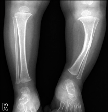
Figure 1 Anteroposterior radiograph of the lower legs demonstrates anterolateral bowing of the left tibia.
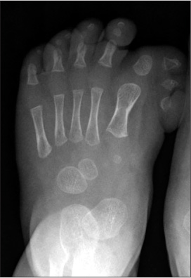
Figure 2 Anteroposterior radiograph of the foot shows duplication of the distal phalanx of the hallux.
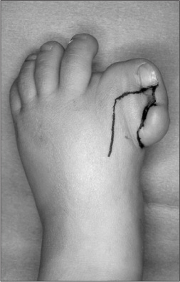
Figure 3 Photograph showing the hallux polydactyly and varus deformity. A proximally based dorsal skin flap is shown.
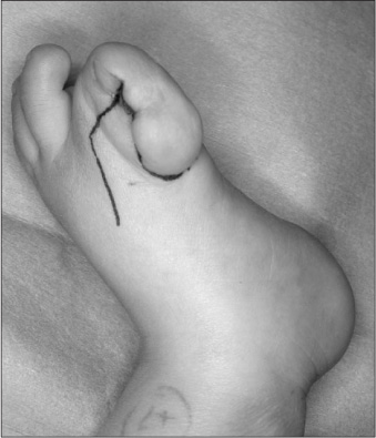
Figure 4 A distally based medial skin flap is designed and the skin flap is defatted.
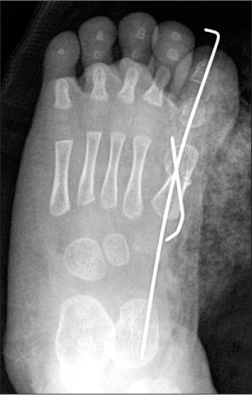
Figure 5 Postoperative radiograph demonstrates metatarsal osteotomy and fixation with K-wires.
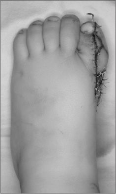
Figure 6 Immediate postoperative photograph the foot.
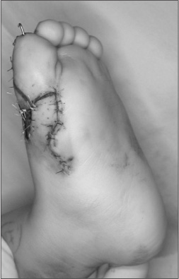
Figure 7 The proximally based dorsal skin flap is translocated into the medial skin defect which had occurred after excision of the extradigit and correction of the varus.
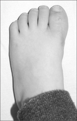
Figure 8 Photograph taken one and a half year postoperatively shows satisfactory correction of the deformity.
Reference
-
1. Canale ST, Beaty JH. Campbell's operative orthopaedics. 2008. 11th ed. Philadelphia: Mosby Elsevier;1063.2. Phelps DA, Grogan DP. Polydactyly of the foot. J Pediatr Orthop. 1985. 5:446–451.
Article3. Venn-Watson EA. Problems in polydactyly of the foot. Orthop Clin North Am. 1976. 7:909–927.
Article4. Watanabe H, Fujita S, Oka I. Polydactyly of the foot: an analysis of 265 cases and a morphological classification. Plast Reconstr Surg. 1992. 89:856–877.5. Masada K, Tsuyuguchi Y, Kawabata H, Ono K. Treatment of preaxial polydactyly of the foot. Plast Reconstr Surg. 1987. 79:251–258.
Article6. Choi IH, Chung CY, Cho TJ, Yoo WJ, Park MS. Duk Yong Lee's pediatric orthopaedics. 2009. 3rd ed. Seoul: Koonja;339.7. Turra S, Gigante C, Bisinella G. Polydactyly of the foot. J Pediatr Orthop B. 2007. 16:216–220.
Article8. Farmer AW. Congenital hallux varus. Am J Surg. 1958. 95:274–278.
Article9. Toriyama K, Kamei Y, Morishita T, Matsuoka K, Torii S. Z-plasty of dorsal and plantar flaps for hallux varus with preaxial polydactyly of the foot. Plast Reconstr Surg. 2006. 117:112e–115e.
Article10. Kardon NB, Dana LP, FitzGerald JM, Opitz JM. Two sporadic cases of amelia/phocomelia with similar phenotype: rare and unusually symmetrical form of FFU dysostosis or separate entity? Am J Med Genet Suppl. 1986. 2:239–245.
Article11. Adamsbaum C, Kalifa G, Seringe R, Bonnet JC. Minor tibial duplication: a new cause of congenital bowing of the tibia. Pediatr Radiol. 1991. 21:185–188.
Article12. Manner HM, Radler C, Ganger R, Grossbötzl G, Petje G, Grill F. Pathomorphology and treatment of congenital anterolateral bowing of the tibia associated with duplication of the hallux. J Bone Joint Surg Br. 2005. 87:226–230.
Article13. Bressers MM, Castelein RM. Anterolateral tibial bowing and duplication of the hallux: a rare but distinct entity with good prognosis. J Pediatr Orthop B. 2001. 10:153–157.
Article14. Lemire EG. Congenital anterolateral tibial bowing and polydactyly: a case report. J Med Case Reports. 2007. 1:54.
Article15. Kitoh H, Nogami H, Hattori T. Congenital anterolateral bowing of the tibia with ipsilateral polydactyly of the great toe. Am J Med Genet. 1997. 73:404–407.
Article16. Weaver KM, Henry GW, Reinker KA. Unilateral duplication of the great toe with anterolateral tibial bowing. J Pediatr Orthop. 1996. 16:73–77.
Article17. Temtamy SA, McKusick VA. The genetics of hand malformations. 1978. New York: Alan R Liss, Inc.;364–372.18. Blauth W, Olason AT. Classification of polydactyly of the hands and feet. Arch Orthop Trauma Surg. 1988. 107:334–344.
- Full Text Links
-
- Actions
-
Cited
- CITED
-
- Close
- Share
-
- Similar articles
-
- Minimally Invasive Surgery with Tenorrhaphy for Postoperative Hallux Varus Deformity Combined with Flexor Hallucis Longus Rupture after Hallux Valgus Correction: A Case Report
- Surgical Treatment of Congenital Hallux Varus
- Simple Classification of Foot Polydactyly Based on The Status of Metatarsal Bone and Varus Deformity
- Complications after Surgical Correction of Hallux Valgus
- Tibial Torsion and Knee Varus Angle: Are These Aetiologies of Hallux Valgus?
