Multi-Detector CT Findings of Double Retroaortic Left Renal Veins
- Affiliations
-
- 1Department of Radiology, Uijeongbu St. Mary's Hospital, College of Medicine, The Catholic University of Korea, Uijeongbu, Korea. ymiku@catholic.ac.kr
- KMID: 1439539
- DOI: http://doi.org/10.3348/jksr.2012.67.4.269
Abstract
- The awareness of renal vascular anomalies is important in order to avoid diagnostic pitfalls and to plan for preoperative surgery and interventional radiology. Retroaortic left renal vein is an uncommon congenital venous variation that is classified type 1 and type 2. However, coexistence of type 1 and type 2, called double retroaortic left renal veins, is an extremely rare variation. Only a few cases of double retroaortic left renal veins have been reported. We present a case of a 51-year-old woman with double retroaortic left renal veins with multi-detector CT findings and review of embryological basis.
Figure
-
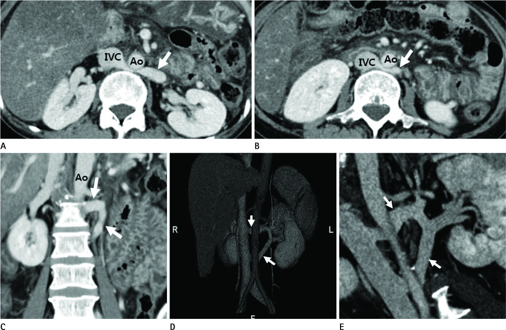
Fig. 1 Contrast-enhanced MDCT images of a 51-year-old woman with double retroaortic left renal veins. Axial CT image at level of L1-2 vertebra (A) shows the left retroaortic renal vein coursing behind the abdominal aorta and draining into the IVC (white arrow). Axial CT image at level of L3-4 vertebra (B) reveals another left retroaortic renal vein that runs dorsally to the abdominal aorta and draining into the IVC (white arrow). Coronal reformatted CT image (C) demonstrates two left retroaortic renal veins. Volume Rendering 3D reconstruction image (D) and Curved multiplanar reformation image (E) show two left retroaortic renal veins coursing behind the abdominal aorta and separately draining into the IVC behind the abdominal aorta. Note.-Ao = aorta, IVC = inferior vena cava, MDCT = multi-detector CT, 3D = three-dimensional

Fig. 2 Conceptual framework for the development of double retroaortic renal veins. A. Coronal diagram shows embryological development of the renal veins and inferior vena cava. Three paired embryonic vessels and their anastomoses contribute the inferior vena cava and renal veins. The subcardinal and the supracardinal veins are interconnected by a network of veins, forming a venous collar around the aorta. B. Coronal diagram shows developmental process of retroaortic left renal vein. Ventral intersubcardinal veins are regressed and remained dorsal intersupracardinal anastomosis and left renal collar veins are formed left renal vein. Regressed embryonic veins are illustrated by dash-line. C. Coronal diagram shows coexistent two types of retroaortic left renal veins in case of our patient. Note.-a = subcardinal vein, b = supracardinal vein, c = postcardinal vein, d, d' = supra-subcardinal anastomosis, renal collar, e = intersubcardinal anastomosis ventral to the aorta, f, f' = intersupracardinal anastomosis dorsal to the aorta, g, g' = renal vein from renal collar, h = post-supracardinal anastomosis, i = post-subcardinal anastomosis, IVC = inferior vena cava
Reference
-
1. Bass JE, Redwine MD, Kramer LA, Huynh PT, Harris JH Jr. Spectrum of congenital anomalies of the inferior vena cava: cross-sectional imaging findings. Radiographics. 2000. 20:639–652.2. Moore KL, Persaud TVN. The cardiovascular system. The developing human: clinically oriented embryology. 2008. 8th ed. London: Saunders;285–336.3. Mathews R, Smith PA, Fishman EK, Marshall FF. Anomalies of the inferior vena cava and renal veins: embryologic and surgical considerations. Urology. 1999. 53:873–880.4. Mayo J, Gray R, St Louis E, Grosman H, McLoughlin M, Wise D. Anomalies of the inferior vena cava. AJR Am J Roentgenol. 1983. 140:339–345.5. Aljabri B, MacDonald PS, Satin R, Stein LS, Obrand DI, Steinmetz OK. Incidence of major venous and renal anomalies relevant to aortoiliac surgery as demonstrated by computed tomography. Ann Vasc Surg. 2001. 15:615–618.6. Koc Z, Ulusan S, Tokmak N, Oguzkurt L, Yildirim T. Double retroaortic left renal veins as a possible cause of pelvic congestion syndrome: imaging findings in two patients. Br J Radiol. 2006. 79:e152–e155.7. Hoeltl W, Hruby W, Aharinejad S. Renal vein anatomy and its implications for retroperitoneal surgery. J Urol. 1990. 143:1108–1114.8. Satyapal KS, Kalideen JM, Haffejee AA, Singh B, Robbs JV. Left renal vein variations. Surg Radiol Anat. 1999. 21:77–81.9. Kara E, Oztürk NC, Ozgür A, Yıldız A, Oztürk H. Ectopic kidney with varied vasculature: demonstrated by CT angiography. Surg Radiol Anat. 2011. 33:81–84.
- Full Text Links
-
- Actions
-
Cited
- CITED
-
- Close
- Share
-
- Similar articles
-
- The Incidence of Inferior Vena Cava Anomalies by MDCT
- Retroaortic Renal Vein
- Posterior Nutcracker Syndrome Associated with Interrupted Left Inferior Vena Cava with Azygos Continuation and Retroaortic Right Renal Vein
- MDCT Findings of Right Circumaortic Renal Vein with Ectopic Kidney
- CT Angiography for Living Kidney Donors: Accuracy, Cause of Misinterpretation and Prevalence of Variation
