Posterior Nutcracker Syndrome Associated with Interrupted Left Inferior Vena Cava with Azygos Continuation and Retroaortic Right Renal Vein
- Affiliations
-
- 1Department of Ultrasonography, Xijing Hospital, Fourth Military Medical University, Shaanxi 710032, China. zhouxiaodong6@gmail.com
- 2Department of Ultrasonography, Fuzhou General Hospital, Fujian 350025, China.
- 3Department of Radiology, Fuzhou General Hospital, Fujian 350025, China.
- KMID: 1372855
- DOI: http://doi.org/10.3348/kjr.2012.13.3.345
Abstract
- Various anatomic anomalies have been considered the causes of nutcracker syndrome (NCS). Posterior NCS refers to the condition, in which vascular narrowing was secondary to the compression of the retroaortic left renal vein while it is crossing between the aorta and the vertebral column. Here, we report an unusual case of posterior NCS associated with a complicated malformation of the interrupted left inferior vena cava with azygos continuation and retroaortic right renal vein, diagnosed by both color Doppler ultrasonography and CT angiography.
MeSH Terms
Figure
-
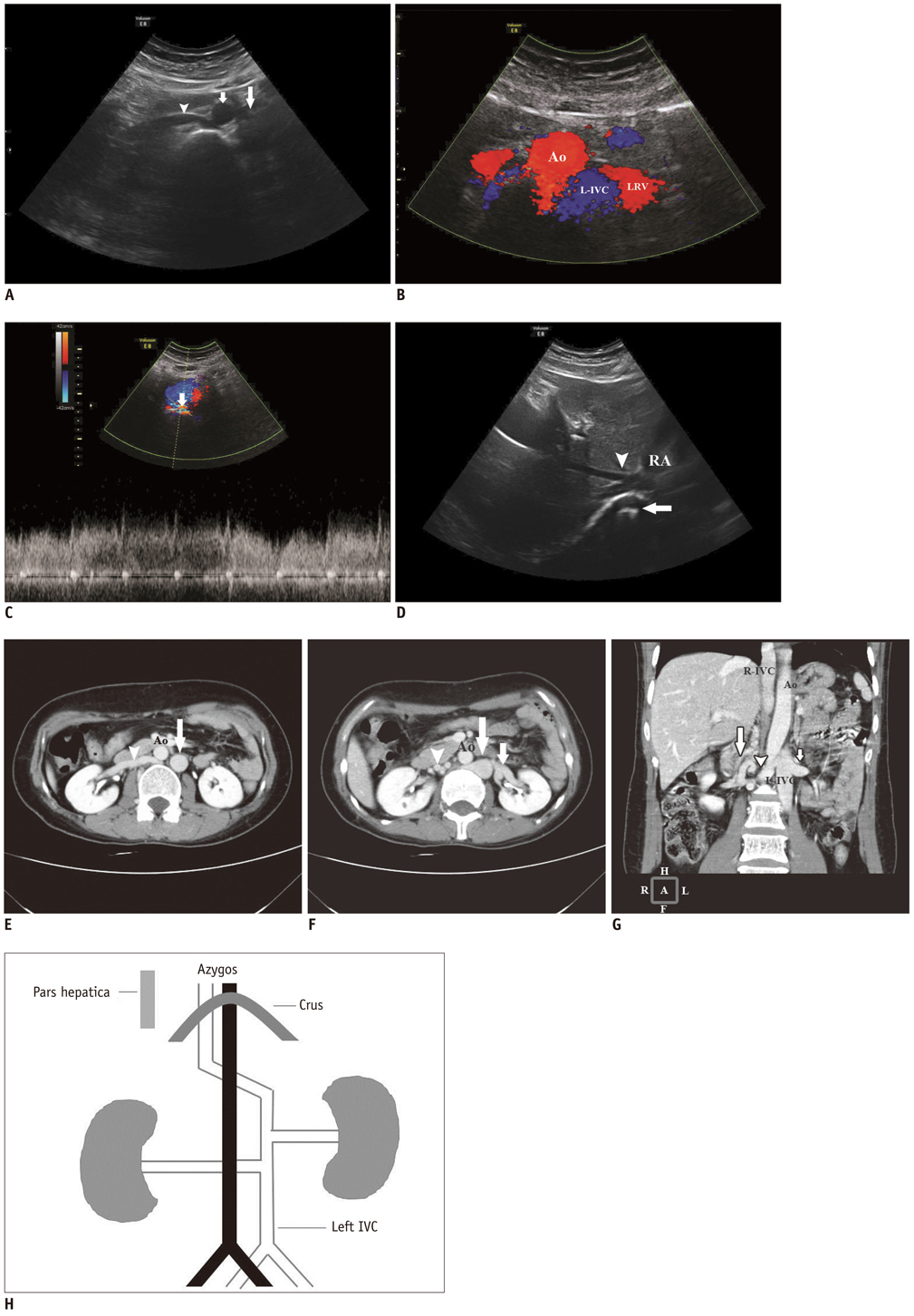
Fig. 1 Complicated malformation of left inferior vena cava presenting as nutcracker syndrome in 42-year-old woman. Axial sonogram shows (A) right renal vein (RRV) (arrowhead) crossing retroaortically to area of left-sided infrarenal inferior vena cava (L-IVC) (long arrow) and no tubular structure parallel to aorta (short arrow) at right side, and (B) two tubular structures at left of aorta at level of left renal vein (LRV). (C) Spectral Doppler sonogram shows the narrowing portion of L-IVC (short arrow) with turbulent high-velocity blood flow between aorta and vertebral column. (D) Axial sonogram reveals that hepatic veins (arrowhead) do not flow to right-sided suprarenal inferior vena cava (R-IVC) (long arrow) but run to right atrium (RA). Ao = aorta Axial CT angiogram is performed and demonstrates (E) right renal vein (RRV) (arrowhead) crossing retroaortically to left-sided infrarenal inferior vena cava (L-IVC) (long arrow) and (F) dilatation of both L-IVC (long arrow) and distal left renal vein (LRV) (short arrow), with the narrowing portion of RRV between aorta and lumber vertebral column. Tortuous paravertebral collateral veins can be seen in right lumbar area (arrowhead). (G) Coronal oblique image shows a dilated L-IVC receiving both LRV (short arrow) and RRV (arrowhead) and running to R-IVC retroaortically. The tortuously paravertebral collateral veins in right lumbar region are also seen (long arrow). (H) Schematic anatomy illustration shows multiple anomalies with the LIVC. Ao = aorta, R-IVC = right-sided suprarenal inferior vena cava
Reference
-
1. de Schepper A. ["Nutcracker" phenomenon of the renal vein and venous pathology of the left kidney]. J Belge Radiol. 1972. 55:507–511.2. Beinart C, Sniderman KW, Saddekni S, Weiner M, Vaughan ED Jr, Sos TA. Left renal vein hypertension: a cause of occult hematuria. Radiology. 1982. 145:647–650.3. Hartung O, Barthelemy P, Berdah SV, Alimi YS. Laparoscopy-assisted left ovarian vein transposition to treat one case of posterior nutcracker syndrome. Ann Vasc Surg. 2009. 23:413.e13–413.e16.4. Fitoz S, Yalcinkaya F. Compression of left inferior vena cava: a form of nutcracker syndrome. J Clin Ultrasound. 2008. 36:101–104.5. Ulusan S, Koc Z. Left inferior vena cava associated with nutcracker phenomenon. Firat Tip Dergisi. 2007. 12:151–153.6. Gupta A, Naik N, Gulati GS. Mesoaortic entrapment of a left inferior vena cava. Indian J Radiol Imaging. 2010. 20:63–65.7. Bass JE, Redwine MD, Kramer LA, Huynh PT, Harris JH Jr. Spectrum of congenital anomalies of the inferior vena cava: cross-sectional imaging findings. Radiographics. 2000. 20:639–652.8. Wong HI, Chen MC, Wu CS, Fu KA, Lin CH, Weng MJ, et al. The usefulness of fast-spin-echo T2-weighted MR imaging in Nutcracker syndrome: a case report. Korean J Radiol. 2010. 11:373–377.9. Kim SH, Cho SW, Kim HD, Chung JW, Park JH, Han MC. Nutcracker syndrome: diagnosis with Doppler US. Radiology. 1996. 198:93–97.10. Pollock C, Liu PL, Györy AZ, Grigg R, Gallery ED, Caterson R, et al. Dysmorphism of urinary red blood cells--value in diagnosis. Kidney Int. 1989. 36:1045–1049.
- Full Text Links
-
- Actions
-
Cited
- CITED
-
- Close
- Share
-
- Similar articles
-
- Absence of the Intrahepatic Inferior Vena Cava with Polysplenia Syndrome on Multidetector Computed Tomography: A Case Report
- A Case of Congenital Absence of the Inferior Vena Cava
- A case of hemiazygos continuation of a left inferior vena cava
- Congenital Interruption of the Inferior Vena Cava with Azygos Continuation: A Case Report
- Congenital Absence of the Azygos Vein with Persistent Left Superior Vena Cava: A Case Report
