Multiparameter Flow Cytometry: Advances in High Resolution Analysis
- Affiliations
-
- 1Research and Development, BD Biosciences, La Jolla, CA 92037, USA. Ravi_Hingorani@BD.COM
- KMID: 1431925
- DOI: http://doi.org/10.4110/in.2013.13.2.43
Abstract
- Over the past 40 years, flow cytometry has emerged as a leading, application-rich technology that supports high-resolution characterization of individual cells which function in complex cellular networks such as the immune system. This brief overview highlights advances in multiparameter flow cytometric technologies and reagent applications for characterization and functional analysis of cells modulating the immune network. These advances significantly support high-throughput and high-content analyses and enable an integrated understanding of the cellular and molecular interactions that underlie complex biological systems.
Keyword
MeSH Terms
Figure
-
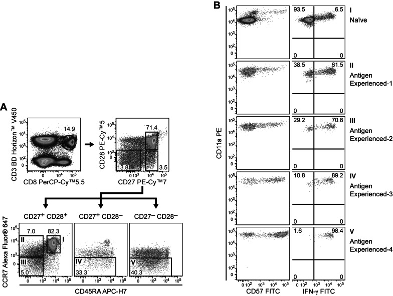
Figure 1 Polychromatic flow cytometry for analysis of T-cell phenotype and effector function. PBMCs were stained with CD57 FITC, CD11a PE, CD28 PE-Cy™5, CD27 PE-Cy™7, CD8 PerCP-Cy™5.5, CCR7 Alexa Fluor® 647, CD45RA APC-H7, CD3 BD Horizon™ V450, and CD4 BD Horizon™ V500. Cells were then acquired on a BD™ LSR II and analyzed for CD3 and for the subsets within the CD8 T-cell population. These subsets were further analyzed based on CD28 and CD27 staining to obtain the CD27+CD28+, CD27+CD28-, and CD27+CD28- fractions as shown in Panel A. Based on CD197 (CCR7) and CD45RA staining, the cells were then identified as Naïve, Antigen experienced-1, Antigen experienced-2, Antigen experienced-3, and Antigen experienced-4 (Panel B) as described in Appay, et al. (2008). Note that the expression of CD57-positive cells increased with the increased antigen experience. To confirm the increase in antigen experience, CD8 subsets from PBMCs were analyzed after stimulation with PMA/Ionomycin in the presence of BD GolgiStop™ protein transport inhibitor for 5 hours. As expected, there was an increase in the intracellular stain for IFN-γ as the cells moved from the naïve phenotype to the more experienced phenotype.
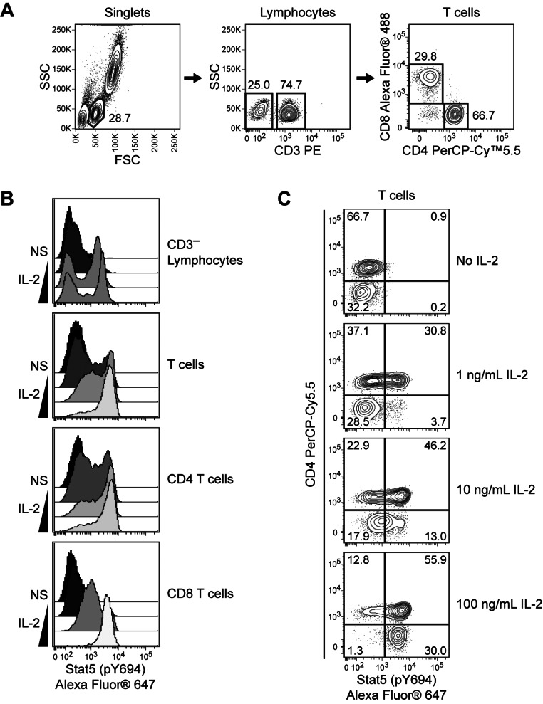
Figure 2 Detection of heterogeneous signaling responses within CD4 and CD8 T-cell populations. Human whole blood was stimulated with 0, 1, 10, or 100 ng/ml of recombinant human IL-2 for 15 minutes at 37℃ and fixed, permeabilized, and stained using the BD Phosflow™ human T cell activation kit. Data was acquired on a BD FACSVerse™ flow cytometer and analyzed using Cytobank software. Lymphocyte subpopulations were identified based on surface marker expression (Panel A), and Stat5 (pY694) phosphorylation responses were assessed within each subpopulation (Panel B) or within T cells (Panel C). IL-2 induced a dose-dependent increase in Stat5 (pY694) phosphorylation in T cells and a subpopulation of CD3- lymphocytes. Compared to CD8 T cells, a larger subpopulation of CD4 T cells responded at the lowest concentration of IL-2. However, a subpopulation of CD4 T cells remained unresponsive to IL-2 at the highest concentration, whereas the majority of CD8 T cells responded strongly.
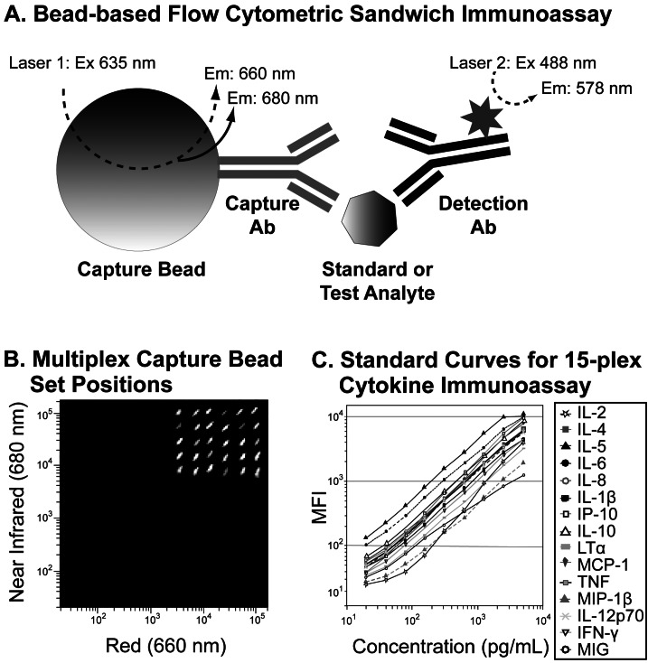
Figure 3 Multiplex flow cytometric immunoassays. Bead-based multiplex immunoassay principles are demonstrated using the BD™ Cytometric Bead Array (CBA) Flex Set system (Panel A). Each set of capture beads is labeled with two different fluorescent dyes and conjugated to a capture antibody specific for a particular analyte. When mixed with test samples or calibrated standards, the capture beads specifically bind and localize standard or test analytes to their surfaces. PE-labeled detection antibodies bind to another site on the analyte. Excitation of capture bead dyes by the red (635 nm) laser allows the identification of each set of capture beads based on its unique fluorescence intensities in the red (660 nm) and near-infrared (680 nm) channels. Beads with different two-color fluorescence positions can be combined to create relatively high content multiplex assays, such as the 30-plex assay shown (Panel B). Blue-laser (488 nm) excitation of the PE-labeled detection antibodies produces signal intensities commensurate with the amount of bound analyte. The flow cytometric data for each capture bead set can be analyzed to generate standard curves and to quantify the levels of specific analytes in test samples. Standard curves generated from a 15-plex Cytokine BD CBA Flex Set analysis are shown (Panel C).
Reference
-
1. Shapiro HM. Practical flow cytometry. 2003. 4th ed. Hoboken, NJ: Wiley-Liss.2. Henel G, Schmitz JL. Basic theory and clinical applications of flow cytometry. Lab Med. 2007; 38:428–436.
Article3. Hayashi T, Shibata N, Okumura R, Kudome T, Nishimura O, Tarui H, Agata K. Single-cell gene profiling of planarian stem cells using fluorescent activated cell sorting and its "index sorting" function for stem cell research. Dev Growth Differ. 2010; 52:131–144. PMID: 20078655.
Article4. Doležel J, Vrána J, Safář J, Bartoš J, Kubaláková M, Simková H. Chromosomes in the flow to simplify genome analysis. Funct Integr Genomics. 2012; 12:397–416. PMID: 22895700.
Article5. Tighe P, Negm O, Todd I, Fairclough L. Utility, reliability and reproducibility of immunoassay multiplex kits. Methods. 2013; [Epub ahead of print].
Article6. Ernst D, Bolton G, Recktenwald D, Cameron MJ, Danesh A, Persad D, Kelvin DJ, Gaur A. Tang YW, Stratton CW, editors. Bead-based flow cytometric assays: A multiplex assay platform with applications in diagnostic microbiology. Advanced techniques in diagnostic microbiology. 2006. New York, U.S.A.: Springer;p. 427–443.
Article7. Morgan E, Varro R, Sepulveda H, Ember JA, Apgar J, Wilson J, Lowe L, Chen R, Shivraj L, Agadir A, Campos R, Ernst D, Gaur A. Cytometric bead array: a multiplexed assay platform with applications in various areas of biology. Clin Immunol. 2004; 110:252–266. PMID: 15047203.
Article8. Funato Y, Baumhover H, Grantham-Wright D, Wilson J, Ernst D, Sepulveda H. Simultaneous measurement of six human cytokines using the Cytometric Bead Array System, a multiparameter immunoassay system for flow cytometry. Cytometry Res. 2002; 12:93–97.9. Cook EB, Stahl JL, Lowe L, Chen R, Morgan E, Wilson J, Varro R, Chan A, Graziano FM, Barney NP. Simultaneous measurement of six cytokines in a single sample of human tears using microparticle-based flow cytometry: allergics vs. non-allergics. J Immunol Methods. 2001; 254:109–118. PMID: 11406157.
Article10. Chattopadhyay PK, Roederer M. Cytometry: today's technology and tomorrow's horizons. Methods. 2012; 57:251–258. PMID: 22391486.
Article11. Herzenberg LA, Parks D, Sahaf B, Perez O, Roederer M, Herzenberg LA. The history and future of the fluorescence activated cell sorter and flow cytometry: a view from Stanford. Clin Chem. 2002; 48:1819–1827. PMID: 12324512.
Article12. Zhang P, Zhao N, Zeng Z, Chang CC, Zu Y. Combination of an aptamer probe to CD4 and antibodies for multicolored cell phenotyping. Am J Clin Pathol. 2010; 134:586–593. PMID: 20855639.
Article13. Telford WG, Hawley T, Subach F, Verkhusha V, Hawley RG. Flow cytometry of fluorescent proteins. Methods. 2012; 57:318–330. PMID: 22293036.
Article14. Robinson JP, Rajwa B, Patsekin V, Davisson VJ. Computational analysis of high-throughput flow cytometry data. Expert Opin Drug Discov. 2012; 7:679–693. PMID: 22708834.
Article15. De Rosa SC, Brenchley JM, Roederer M. Beyond six colors: a new era in flow cytometry. Nat Med. 2003; 9:112–117. PMID: 12514723.
Article16. Appay V, van Lier RA, Sallusto F, Roederer M. Phenotype and function of human T lymphocyte subsets: consensus and issues. Cytometry A. 2008; 73:975–983. PMID: 18785267.
Article17. Craig FE, Foon KA. Flow cytometric immunophenotyping for hematologic neoplasms. Blood. 2008; 111:3941–3967. PMID: 18198345.
Article18. Danna EA, Nolan GP. Transcending the biomarker mindset: deciphering disease mechanisms at the single cell level. Curr Opin Chem Biol. 2006; 10:20–27. PMID: 16406766.
Article19. Krutzik PO, Crane JM, Clutter MR, Nolan GP. High-content single-cell drug screening with phosphospecific flow cytometry. Nat Chem Biol. 2008; 4:132–142. PMID: 18157122.
Article20. Maino VC, Picker LJ. Identification of functional subsets by flow cytometry: intracellular detection of cytokine expression. Cytometry. 1998; 34:207–215. PMID: 9822306.
Article21. Perez OD, Mitchell D, Campos R, Gao GJ, Li L, Nolan GP. Multiparameter analysis of intracellular phosphoepitopes in immunophenotyped cell populations by flow cytometry. Curr Protoc Cytom. 2005; Chapter 6:Unit 6.20. PMID: 18770823.
Article22. Chattopadhyay PK, Gaylord B, Palmer A, Jiang N, Raven MA, Lewis G, Reuter MA, Nur-ur Rahman AK, Price DA, Betts MR, Roederer M. Brilliant violet fluorophores: a new class of ultrabright fluorescent compounds for immunofluorescence experiments. Cytometry A. 2012; 81:456–466. PMID: 22489009.
Article23. Chattopadhyay PK. Quantum dot technology in flow cytometry. Methods Cell Biol. 2011; 102:463–477. PMID: 21704850.
Article24. Kalina T, Flores-Montero J, van der Velden VH, Martin-Ayuso M, Böttcher S, Ritgen M, Almeida J, Lhermitte L, Asnafi V, Mendonça A, de Tute R, Cullen M, Sedek L, Vidriales MB, Pérez JJ, te Marvelde JG, Mejstrikova E, Hrusak O, Szczepański T, van Dongen JJ, Orfao A. EuroFlow Consortium (EU-FP6, LSHB-CT-2006-018708). EuroFlow standardization of flow cytometer instrument settings and immunophenotyping protocols. Leukemia. 2012; 26:1986–2010. PMID: 22948490.
Article25. Crissman HA, Darzynkiewicz Z, Tobey RA, Steinkamp JA. Correlated measurements of DNA, RNA, and protein in individual cells by flow cytometry. Science. 1985; 228:1321–1324. PMID: 2408339.
Article26. Peixoto A, Monteiro M, Rocha B, Veiga-Fernandes H. Quantification of multiple gene expression in individual cells. Genome Res. 2004; 14:1938–1947. PMID: 15466292.
Article27. Krutzik PO, Clutter MR, Trejo A, Nolan GP. Fluorescent cell barcoding for multiplex flow cytometry. Curr Protoc Cytom. 2011; Chapter 6:Unit 6.31. PMID: 21207359.
Article28. Krutzik PO, Nolan GP. Fluorescent cell barcoding in flow cytometry allows high-throughput drug screening and signaling profiling. Nat Methods. 2006; 3:361–368. PMID: 16628206.
Article29. Bodenmiller B, Zunder ER, Finck R, Chen TJ, Savig ES, Bruggner RV, Simonds EF, Bendall SC, Sachs K, Krutzik PO, Nolan GP. Multiplexed mass cytometry profiling of cellular states perturbed by small-molecule regulators. Nat Biotechnol. 2012; 30:858–867. PMID: 22902532.
Article30. Nomura L, Maino VC, Maecker HT. Standardization and optimization of multiparameter intracellular cytokine staining. Cytometry A. 2008; 73:984–991. PMID: 18612990.
Article31. Suni MA, Dunn HS, Orr PL, de Laat R, Sinclair E, Ghanekar SA, Bredt BM, Dunne JF, Maino VC, Maecker HT. Performance of plate-based cytokine flow cytometry with automated data analysis. BMC Immunol. 2003; 4:9. PMID: 12952557.32. Gray JW, Darzynkiewicz Z. Techniques in cell cycle analysis. 1987. Totowa, NJ: Humana Press.33. Darzynkiewicz Z, Traganos F, Sharpless T, Melamed MR. Lymphocyte stimulation: a rapid multiparameter analysis. Proc Natl Acad Sci U S A. 1976; 73:2881–2884. PMID: 822422.
Article34. Cappella P, Gasparri F, Pulici M, Moll J. A novel method based on click chemistry, which overcomes limitations of cell cycle analysis by classical determination of BrdU incorporation, allowing multiplex antibody staining. Cytometry A. 2008; 73:626–636. PMID: 18521918.
Article35. Feig C, Peter ME. How apoptosis got the immune system in shape. Eur J Immunol. 2007; 37(Suppl 1):S61–S70. PMID: 17972347.
Article36. Hanahan D, Weinberg RA. Hallmarks of cancer: the next generation. Cell. 2011; 144:646–674. PMID: 21376230.
Article37. Wlodkowic D, Telford W, Skommer J, Darzynkiewicz Z. Apoptosis and beyond: cytometry in studies of programmed cell death. Methods Cell Biol. 2011; 103:55–98. PMID: 21722800.
Article38. Wlodkowic D, Skommer J, Darzynkiewicz Z. Cytometry in cell necrobiology revisited. Recent advances and new vistas. Cytometry A. 2010; 77:591–606. PMID: 20235235.
Article39. Tanaka T, Halicka HD, Traganos F, Seiter K, Darzynkiewicz Z. Induction of ATM activation, histone H2AX phosphorylation and apoptosis by etoposide: relation to cell cycle phase. Cell Cycle. 2007; 6:371–376. PMID: 17297310.
Article40. Schulz KR, Danna EA, Krutzik PO, Nolan GP. Single-cell phospho-protein analysis by flow cytometry. Curr Protoc Immunol. 2012; Chapter 8:Unit 8.17.1–Unit 8.17.20.
Article41. Juan G, Traganos F, James WM, Ray JM, Roberge M, Sauve DM, Anderson H, Darzynkiewicz Z. Histone H3 phosphorylation and expression of cyclins A and B1 measured in individual cells during their progression through G2 and mitosis. Cytometry. 1998; 32:71–77. PMID: 9627219.
Article42. Fleisher TA, Dorman SE, Anderson JA, Vail M, Brown MR, Holland SM. Detection of intracellular phosphorylated STAT-1 by flow cytometry. Clin Immunol. 1999; 90:425–430. PMID: 10075873.
Article43. Chow S, Patel H, Hedley DW. Measurement of MAP kinase activation by flow cytometry using phospho-specific antibodies to MEK and ERK: potential for pharmacodynamic monitoring of signal transduction inhibitors. Cytometry. 2001; 46:72–78. PMID: 11309815.
Article44. Zell T, Khoruts A, Ingulli E, Bonnevier JL, Mueller DL, Jenkins MK. Single-cell analysis of signal transduction in CD4 T cells stimulated by antigen in vivo. Proc Natl Acad Sci U S A. 2001; 98:10805–10810. PMID: 11535838.
Article45. Perez OD, Nolan GP. Simultaneous measurement of multiple active kinase states using polychromatic flow cytometry. Nat Biotechnol. 2002; 20:155–162. PMID: 11821861.
Article46. Tazzari PL, Cappellini A, Bortul R, Ricci F, Billi AM, Tabellini G, Conte R, Martelli AM. Flow cytometric detection of total and serine 473 phosphorylated Akt. J Cell Biochem. 2002; 86:704–715. PMID: 12210737.
Article47. Krutzik PO, Nolan GP. Intracellular phospho-protein staining techniques for flow cytometry: monitoring single cell signaling events. Cytometry A. 2003; 55:61–70. PMID: 14505311.
Article48. Chow S, Hedley D, Grom P, Magari R, Jacobberger JW, Shankey TV. Whole blood fixation and permeabilization protocol with red blood cell lysis for flow cytometry of intracellular phosphorylated epitopes in leukocyte subpopulations. Cytometry A. 2005; 67:4–17. PMID: 16080188.
Article49. Suni MA, Maino VC. Flow cytometric analysis of cell signaling proteins. Methods Mol Biol. 2011; 717:155–169. PMID: 21370030.
Article50. Irish JM, Czerwinski DK, Nolan GP, Levy R. Altered B-cell receptor signaling kinetics distinguish human follicular lymphoma B cells from tumor-infiltrating nonmalignant B cells. Blood. 2006; 108:3135–3142. PMID: 16835385.
Article51. Sadeghi K, Berger A, Langgartner M, Prusa AR, Hayde M, Herkner K, Pollak A, Spittler A, Forster-Waldl E. Immaturity of infection control in preterm and term newborns is associated with impaired toll-like receptor signaling. J Infect Dis. 2007; 195:296–302. PMID: 17191175.
Article52. Zeiser R, Leveson-Gower DB, Zambricki EA, Kambham N, Beilhack A, Loh J, Hou JZ, Negrin RS. Differential impact of mammalian target of rapamycin inhibition on CD4+CD25+Foxp3+ regulatory T cells compared with conventional CD4+ T cells. Blood. 2008; 111:453–462. PMID: 17967941.
Article53. Perfetto SP, Chattopadhyay PK, Lamoreaux L, Nguyen R, Ambrozak D, Koup RA, Roederer M. Amine-reactive dyes for dead cell discrimination in fixed samples. Curr Protoc Cytom. 2010; Chapter 9:Unit 9.34. PMID: 20578108.
Article54. Henriksen M, Miller B, Newmark J, Al-Kofahi Y, Holden E. Laser scanning cytometry and its applications: a pioneering technology in the field of quantitative imaging cytometry. Methods Cell Biol. 2011; 102:161–205. PMID: 21704839.
Article55. Zuba-Surma EK, Ratajczak MZ. Analytical capabilities of the ImageStream cytometer. Methods Cell Biol. 2011; 102:207–230. PMID: 21704840.
Article56. Clutter MR, Heffner GC, Krutzik PO, Sachen KL, Nolan GP. Tyramide signal amplification for analysis of kinase activity by intracellular flow cytometry. Cytometry A. 2010; 77:1020–1031. PMID: 20824632.
Article57. Krutzik PO, Trejo A, Schulz KR, Nolan GP. Phospho flow cytometry methods for the analysis of kinase signaling in cell lines and primary human blood samples. Methods Mol Biol. 2011; 699:179–202. PMID: 21116984.
Article58. Kuckuck FW, Edwards BS, Sklar LA. High throughput flow cytometry. Cytometry. 2001; 44:83–90. PMID: 11309812.
Article59. Bashashati A, Brinkman RR. A survey of flow cytometry data analysis methods. Adv Bioinformatics. 2009; 584603. PMID: 20049163.
Article60. Lugli E, Roederer M, Cossarizza A. Data analysis in flow cytometry: the future just started. Cytometry A. 2010; 77:705–713. PMID: 20583274.
Article61. Aghaeepour N, Nikolic R, Hoos HH, Brinkman RR. Rapid cell population identification in flow cytometry data. Cytometry A. 2011; 79:6–13. PMID: 21182178.
Article62. Sachs K, Gentles AJ, Youland R, Itani S, Irish J, Nolan GP, Plevritis SK. Characterization of patient specific signaling via augmentation of Bayesian networks with disease and patient state nodes. Conf Proc IEEE Eng Med Biol Soc. 2009; 2009:6624–6627. PMID: 19963681.
Article63. Qiu P, Simonds EF, Bendall SC, Gibbs KD Jr, Bruggner RV, Linderman MD, Sachs K, Nolan GP, Plevritis SK. Extracting a cellular hierarchy from high-dimensional cytometry data with SPADE. Nat Biotechnol. 2011; 29:886–891. PMID: 21964415.
Article64. Pedreira CE, Costa ES, Barrena S, Lecrevisse Q, Almeida J, van Dongen JJ, Orfao A. EuroFlow Consortium. Generation of flow cytometry data files with a potentially infinite number of dimensions. Cytometry A. 2008; 73:834–846. PMID: 18629843.
Article65. Bagwell CB. Breaking the dimensionality barrier. Methods Mol Biol. 2011; 699:31–51. PMID: 21116977.
Article66. Bendall SC, Simonds EF, Qiu P, Amir el AD, Krutzik PO, Finck R, Bruggner RV, Melamed R, Trejo A, Ornatsky OI, Balderas RS, Plevritis SK, Sachs K, Pe'er D, Tanner SD, Nolan GP. Single-cell mass cytometry of differential immune and drug responses across a human hematopoietic continuum. Science. 2011; 332:687–696. PMID: 21551058.
Article67. Ornatsky O, Bandura D, Baranov V, Nitz M, Winnik MA, Tanner S. Highly multiparametric analysis by mass cytometry. J Immunol Methods. 2010; 361:1–20. PMID: 20655312.
Article68. Auksorius E, Bromberg Y, Motiejúnaité R, Pieretti A, Liu L, Coron E, Aranda J, Goldstein AM, Bouma BE, Kazlauskas A, Tearney GJ. Dual-modality fluorescence and full-field optical coherence microscopy for biomedical imaging applications. Biomed Opt Express. 2012; 3:661–666. PMID: 22435110.
Article69. Barteneva NS, Fasler-Kan E, Vorobjev IA. Imaging flow cytometry: coping with heterogeneity in biological systems. J Histochem Cytochem. 2012; 60:723–733. PMID: 22740345.70. Pierzchalski A, Mittag A, Tárnok A. Introduction A: recent advances in cytometry instrumentation, probes, and methods--review. Methods Cell Biol. 2011; 102:1–21. PMID: 21704833.71. Shapiro HM, Perlmutter NG. Personal cytometers: slow flow or no flow? Cytometry A. 2006; 69:620–630. PMID: 16680701.
Article72. Petersen TW, Brent Harrison C, Horner DN, van den Engh G. Flow cytometric characterization of marine microbes. Methods. 2012; 57:350–358. PMID: 22796378.
Article73. Wlodkowic D, Khoshmanesh K, Akagi J, Williams DE, Cooper JM. Wormometry-on-a-chip: Innovative technologies for in situ analysis of small multicellular organisms. Cytometry A. 2011; 79:799–813. PMID: 21548078.
Article74. Orozco AF, Lewis DE. Flow cytometric analysis of circulating microparticles in plasma. Cytometry A. 2010; 77:502–514. PMID: 20235276.
Article75. Chang Q, Hedley D. Emerging applications of flow cytometry in solid tumor biology. Methods. 2012; 57:359–367. PMID: 22503773.
Article76. Tárnok A, Ulrich H, Bocsi J. Phenotypes of stem cells from diverse origin. Cytometry A. 2010; 77:6–10. PMID: 20024907.77. Ochatt SJ. Flow cytometry in plant breeding. Cytometry A. 2008; 73:581–598. PMID: 18431774.
Article
- Full Text Links
-
- Actions
-
Cited
- CITED
-
- Close
- Share
-
- Similar articles
-
- Analysis of T Cells Using Flow Cytometry
- A Comparative Study of DNA Quantitation by Image Cytometry and Flow Cytometry
- Advances in Automated Urinalysis Systems, Flow Cytometry and Digitized Microscopy
- The Value of Urine Cytology and Flow Cytometry in Superficial Bladder Tumors
- DNA analysis of body cavity fluids using flow cytometry
