Lab Anim Res.
2013 Jun;29(2):127-130. 10.5625/lar.2013.29.2.127.
Uterine leiomyosarcoma in a wild rat (Rattus norvegicus): usefulness of Ki-67 labeling index for diagnosis
- Affiliations
-
- 1College of Veterinary Medicine and Institute of Veterinary Science, Kangwon National University, Chuncheon, Korea. byoon@kangwon.ac.kr
- 2Toxicologic Pathology Divison, Korea Institute of Toxicology, Daejeon, Korea.
- KMID: 1431347
- DOI: http://doi.org/10.5625/lar.2013.29.2.127
Abstract
- Uterine smooth muscle tumor is very rare in laboratory rats and, there has been no report in the wild rodents. Among a total of 400 wild rats captured in Gyeonggi, Gangwon, and Chungbuk provinces of Korea in 2007, 2010, and 2011, we found a uterine spindle cell tumor, diagnosed as smooth muscle cell origin based on differential features of histology and immunohistochemistry. Its incidence was very low, like in the laboratory rats, as under 0.5% for female. Considering generally applied histological and cellular criteria, this case was difficult in differential diagnosis between benign and malignant. Ki-67 labeling index was therefore further investigated, and it ranged from 26.4 to 37.6% in the 10 different areas, representing an average of 32.9+/-0.05%. The Ki-67 labeling index of neoplastic cells near the necrotic area was recorded as 83.5%. According to such high Ki-67 labeling index, it was more likely a malignant leiomyosarcoma, assenting to the previous proposal that Ki-67 labeling index is a significant criterion to differentiate between malignant and benign in the smooth muscle tumors.
Keyword
MeSH Terms
Figure
-
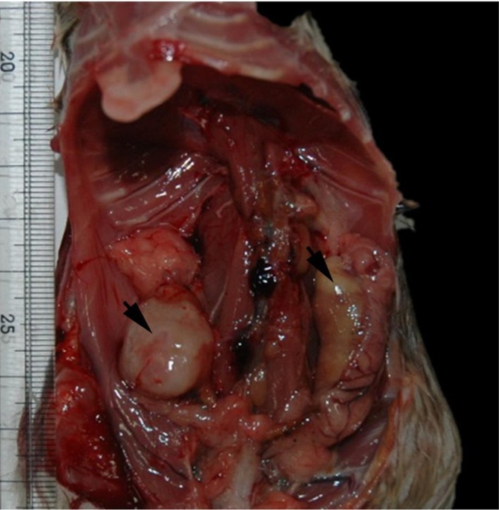
Figure 1 Gross finding of the masses (arrows) in both side of the uterus.
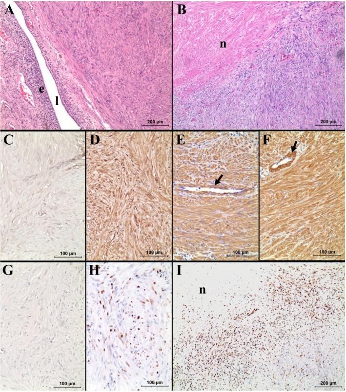
Figure 2 Histopathology (A, B) and immunohistochemistry for cytokeratin (C), vimentin (D), desmin (E), smooth muscle actin (F), S-100 (G) and Ki-67 (H, I). Note the strong immunoreactivity of the neoplastic cells for vimentin, desmin and smooth muscle actin, same as the smooth muscle fibers of blood vessels (arrows in E and F). Also note the very high Ki-67 index of the neoplastic cells near the necrotic area ("n" in B and I). In A, "e" and "l" represent endometrium and lumen, respectively.
Reference
-
1. Cooper BJ, Valentine BA. Tumors of muscles. In : Meuten DJ, editor. Tumor in Domestic Animals. 4th ed. Ames: Iowa State Press;2002. p. 319–363.2. Joel RL, Micheal PJ. Oviduct, Uterus, and Vagina. In : Boorman GA, Eustis SL, Elwell MR, editors. Pathology of the Fischer Rat - Reference and Atlas. San Diego: Academic Press;1990. p. 443–459.3. Bullock FD, Curtis MR. Spontaneous Tumors of the Rat. Cancer Res. 1930; 14:1–115.4. Crain RC. Spontaneous tumors in the Rochester strain of the Wistar rat. Am J Pathol. 1958; 34(2):311–335. PMID: 13520911.5. Yi JY, Kim YH, Yoon BI. Primary subcutaneous leiomyosarcoma of the hamster hind leg. J Vet Med Sci. 2008; 70(5):517–520. PMID: 18525178.
Article6. Anisimov VN, Nikonov AA. Tumours of the vagina, uterus and oviduct. In : Turusov VS, Mohr U, editors. Pathology of tumours in laboratory animals. volume 1. Tumours of the rat. 2nd ed. Oxford: Oxford University Press;1990. p. 445–471.7. Johnson GC, Miller MA, Ramos-Vara JA. Comparison of argyrophilic nucleolar organizer regions (AgNORs) and mitotic index in distinguishing benign from malignant canine smooth muscle tumors and in separating inflammatory hyperplasia from neoplastic lesions of the urinary bladder mucosa. J Vet Diagn Invest. 1995; 7(1):127–136. PMID: 7779947.
Article8. Chen L, Yang B. Immunohistochemical analysis of p16, p53, and Ki-67 expression in uterine smooth muscle tumors. Int J Gynecol Pathol. 2008; 27(3):326–332. PMID: 18580309.
Article9. Mayerhofer K, Lozanov P, Bodner K, Bodner-Adler B, Kimberger O, Czerwenka K. Ki-67 expression in patients with uterine leiomyomas, uterine smooth muscle tumors of uncertain malignant potential (STUMP) and uterine leiomyosarcomas (LMS). Acta Obstet Gynecol Scand. 2004; 83(11):1085–1088. PMID: 15488127.
Article10. Mittal K, Demopoulos RI. MIB-1 (Ki-67), p53, estrogen receptor, and progesterone receptor expression in uterine smooth muscle tumors. Hum Pathol. 2001; 32(9):984–987. PMID: 11567229.
Article11. O'Neill CJ, McBride HA, Connolly LE, McCluggage WG. Uterine leiomyosarcomas are characterized by high p16, p53 and MIB1 expression in comparison with usual leiomyomas, leiomyoma variants and smooth muscle tumours of uncertain malignant potential. Histopathology. 2007; 50(7):851–858. PMID: 17543074.
- Full Text Links
-
- Actions
-
Cited
- CITED
-
- Close
- Share
-
- Similar articles
-
- Prevalence of Capillaria hepatica among house rat in Seoul
- Helminths in Rattus norvegicus captured in Chunchon, Korea
- Expressions and Diagnostic Usefulness of MIB-1 and p53 in Uterine Smooth Muscle Tumors
- Studies on the Expression of the p16 (INK4A), p53, and Ki-67 Labeling Index in Inflammatory and Neoplastic Diseases of the Uterine Cervix
- Expression of Cyclin A and Ki-67 in the Uterine Cervical Carcinoma
