Korean J Radiol.
2012 Dec;13(6):812-815. 10.3348/kjr.2012.13.6.812.
Imaging Finding of Malignant Melanoma of Eustachian Tube with Extension to Middle Ear Cavity: Case Report
- Affiliations
-
- 1Department of Radiology, Yeungnam University College of Medicine, Daegu 705-717, Korea.
- 2Daekyung Radiologic Clinics, Daegu 706-824, Korea.
- 3Department of Radiology, Kyungpook National University Hospital, Daegu 700-721, Korea. leehuijoong@knu.ac.kr
- KMID: 1397514
- DOI: http://doi.org/10.3348/kjr.2012.13.6.812
Abstract
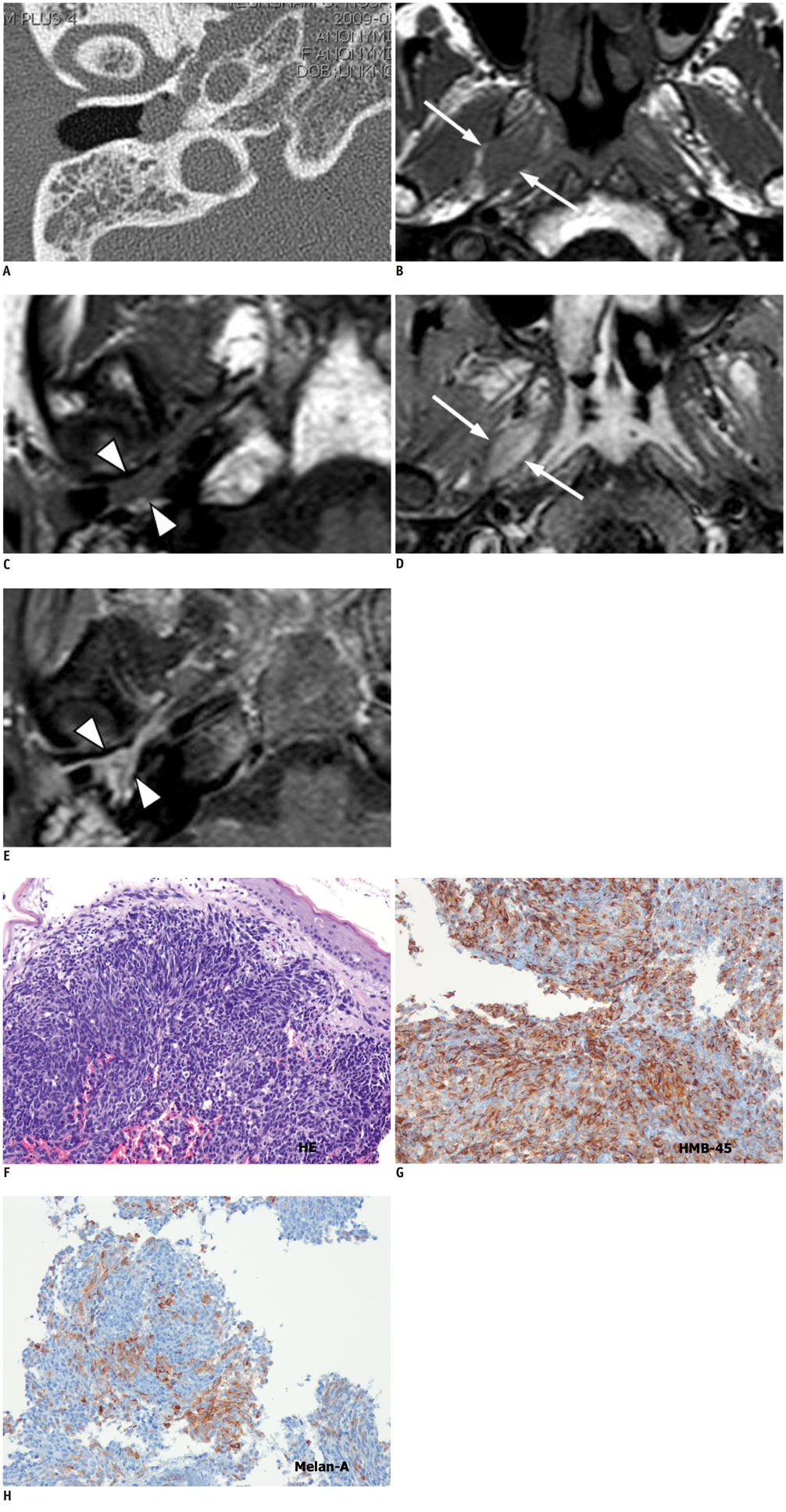
- We report a case of malignant melanoma of Eustachian tube with extension to the middle ear cavity and nasopharynx in a 51-year-old woman who presented with right ear fullness. Computed tomography showed a soft tissue mass in the middle ear cavity and causedthe widening and eroding of the bony eustachian tube. Magnetic resonance imaging showed well enhancing mass in eustachian tube extending nasopharynx to middle ear cavity. A biopsy of the middle ear cavity mass revealed a malignant amelanotic melanoma.
Keyword
MeSH Terms
Figure
Reference
-
1. Lai CC, Tsay SH, Ho CY. Malignant melanoma of the eustachian tube. J Laryngol Otol. 2001. 115:567–569.2. Racic G, Kurtovic D, Roje Z, Tomic S, Dogas Z. Primary mucosal melanoma of the eustachian tube. Eur Arch Otorhinolaryngol. 2004. 261:139–142.3. Baek SJ, Song MH, Lim BJ, Lee WS. Mucosal melanoma arising in the eustachian tube. J Laryngol Otol. 2006. 120:E17.4. Tanaka H, Kohno A, Gomi N, Matsueda K, Mitani H, Kawabata K, et al. Malignant mucosal melanoma of the eustachian tube. Radiat Med. 2008. 26:305–308.5. Yang BT, Wang ZC, Xian JF, Chen QH. MR imaging features of primary mucosal melanoma of the eustachian tube: report of 2 cases. AJNR Am J Neuroradiol. 2009. 30:431–433.6. Batsakis JG, Regezi JA, Solomon AR, Rice DH. The pathology of head and neck tumors: mucosal melanomas, part 13. Head Neck Surg. 1982. 4:404–418.7. Snow GB, van der Waal I. Mucosal melanomas of the head and neck. Otolaryngol Clin North Am. 1986. 19:537–547.8. Uchida M, Matsunami T. Malignant amelanotic melanoma of the middle ear. Arch Otolaryngol Head Neck Surg. 2001. 127:1126–1128.9. Isiklar I, Leeds NE, Fuller GN, Kumar AJ. Intracranial metastatic melanoma: correlation between MR imaging characteristics and melanin content. AJR Am J Roentgenol. 1995. 165:1503–1512.10. Yousem DM, Li C, Montone KT, Montgomery L, Loevner LA, Rao V, et al. Primary malignant melanoma of the sinonasal cavity: MR imaging evaluation. Radiographics. 1996. 16:1101–1110.11. Hirunpat S, Riabroi K, Dechsukhum C, Atchariyasathian V, Tanomkiat W. Nasopharyngeal extension of glomus tympanicum: an unusual clinical and imaging manifestation. AJNR Am J Neuroradiol. 2006. 27:1820–1822.12. Lum C, Keller AM, Kassel E, Blend R, Waldron J, Rutka J. Unusual eustachian tube mass: glomus tympanicum. AJNR Am J Neuroradiol. 2001. 22:508–509.
- Full Text Links
- Actions
-
Cited
- CITED
-
- Close
- Share
- Similar articles
-
- A Case of Eustachian Tube Mature Teratoma
- Luminal development of the eustachian tube and middle ear: murine model
- Comparison of Eustachian Tube Function Before and After Septoplasty: A Systematic Review and Meta-Analysis
- A Case of Middle Ear Effusion due to Lipoma on the Eustachian Tube
- A Case of Nasopharyngeal Carcinoma Presenting as Middle Ear Tumor


