Korean J Radiol.
2012 Dec;13(6):752-759. 10.3348/kjr.2012.13.6.752.
Prognostic Value of Volume-Based 18F-Fluorodeoxyglucose PET/CT Parameters in Patients with Clinically Node-Negative Oral Tongue Squamous Cell Carcinoma
- Affiliations
-
- 1Department of Nuclear Medicine, Ajou University School of Medicine, Suwon 443-721, Korea.
- 2Department of Nuclear Medicine, Samsung Medical Center, Sungkyunkwan University School of Medicine, Seoul 135-710, Korea. jynm.choi@samsung.com
- 3Department of Otorhinolaryngology, Samsung Medical Center, Sungkyunkwan University School of Medicine, Seoul 135-710, Korea.
- KMID: 1397506
- DOI: http://doi.org/10.3348/kjr.2012.13.6.752
Abstract
OBJECTIVE
To evaluate the prognostic value of volume-based metabolic parameters measured with 18F-fluorodeoxyglucose (18F-FDG) positron emission tomography (PET) in patients with clinically node-negative (cN0) oral tongue squamous cell carcinoma (OTSCC) as compared with other prognostic factors.
MATERIALS AND METHODS
In this study, we included a total of 57 patients who had been diagnosed with cN0 tongue cancer by radiologic, 18F-FDG PET/CT, and physical examinations. The maximum standardized uptake value (SUVmax), average SUV (SUVavg), metabolic tumor volume (MTV), and total lesion glycolysis (TLG) for primary tumors were measured with 18F-FDG PET. The prognostic significances of these parameters and other clinical variables were assessed by Cox proportional hazards regression analysis.
RESULTS
In the univariate analysis, pathological node (pN) stage, American Joint Committee on Cancer (AJCC) stage, SUVmax, SUVavg, MTV, and TLG were significant predictors for survival. On a multivariate analysis, pN stage (hazard ratio = 10.555, p = 0.049), AJCC stage (hazard ratio = 13.220, p = 0.045), and MTV (hazard ratio = 2.698, p = 0.033) were significant prognostic factors in cN0 OTSCC patients. The patients with MTV > or = 7.78 cm3 showed a worse prognosis than those with MTV < 7.78 cm3 (p = 0.037).
CONCLUSION
The MTV of primary tumor as a volumetric parameter of 18F-FDG PET, in addition to pN stage and AJCC stage, is an independent prognostic factor for survival in cN0 OTSCC.
Keyword
MeSH Terms
-
Adult
Aged
Carcinoma, Squamous Cell/diagnosis/mortality/pathology/*radionuclide imaging
Female
Fluorodeoxyglucose F18/*diagnostic use
Humans
Lymphatic Metastasis
Magnetic Resonance Imaging
Male
Middle Aged
*Positron-Emission Tomography and Computed Tomography
Prognosis
Radiopharmaceuticals/*diagnostic use
Survival Rate
Tomography, X-Ray Computed
Tongue Neoplasms/diagnosis/mortality/pathology/*radionuclide imaging
Young Adult
Figure
-
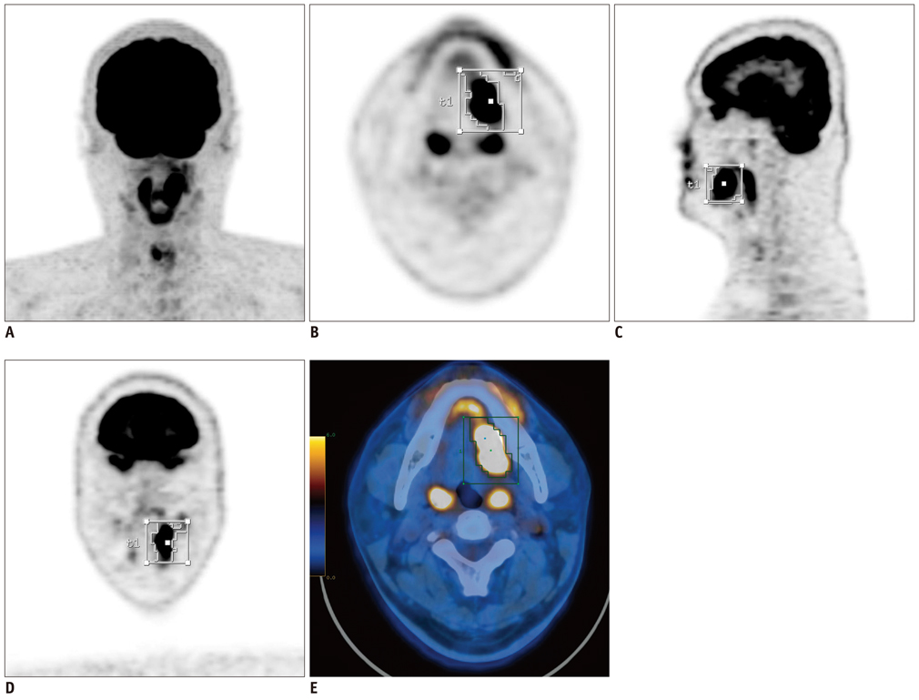
Fig. 1 18F-FDG PET/CT images from 35-year-old male patient with squamous cell carcinoma of tongue. Primary tumor uptake is well visualized on MIP image (A). Segmented VOIs are shown on transverse (B), sagittal (C), coronal (D), and fused PET/CT (E) images. MIP = maximum-intensity-projection, VOI = volume of interest. 18F-FDG PET/CT = 18F-fluorodeoxyglucose positron emission tomography/CT
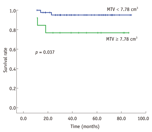
Fig. 2 Kaplan-Meier survival curve according to metabolic tumor volume of primary tumor in clinically node-negative oral tongue squamous cell carcinoma.
Reference
-
1. Bello IO, Soini Y, Salo T. Prognostic evaluation of oral tongue cancer: means, markers and perspectives (I). Oral Oncol. 2010. 46:630–635.2. Bello IO, Soini Y, Salo T. Prognostic evaluation of oral tongue cancer: means, markers and perspectives (II). Oral Oncol. 2010. 46:636–643.3. Regezi JA, Sciubba JJ, Jordan RCK. Oral pathology: Clinical, pathologic correlations. 2007. 5th ed. St. Louis, MO: Saunders Elsevier.4. Leipzig B, Cummings CW, Chung CT, Johnson JT, Sagerman RH. Carcinoma of the anterior tongue. Ann Otol Rhinol Laryngol. 1982. 91(1 Pt 1):94–97.5. Rusthoven K, Ballonoff A, Raben D, Chen C. Poor prognosis in patients with stage I and II oral tongue squamous cell carcinoma. Cancer. 2008. 112:345–351.6. Sathyan KM, Sailasree R, Jayasurya R, Lakshminarayanan K, Abraham T, Nalinakumari KR, et al. Carcinoma of tongue and the buccal mucosa represent different biological subentities of the oral carcinoma. J Cancer Res Clin Oncol. 2006. 132:601–609.7. Zanation AM, Sutton DK, Couch ME, Weissler MC, Shockley WW, Shores CG. Use, accuracy, and implications for patient management of [18F]-2-fluorodeoxyglucose-positron emission/computerized tomography for head and neck tumors. Laryngoscope. 2005. 115:1186–1190.8. Fletcher JW, Djulbegovic B, Soares HP, Siegel BA, Lowe VJ, Lyman GH, et al. Recommendations on the use of 18F-FDG PET in oncology. J Nucl Med. 2008. 49:480–508.9. Shin KM, Lee KS, Shim YM, Kim J, Kim BT, Kwon OJ, et al. FDG PET/CT and mediastinal nodal metastasis detection in stage T1 non-small cell lung cancer: prognostic implications. Korean J Radiol. 2008. 9:481–489.10. Baek CH, Chung MK, Jeong HS, Son YI, Choi J, Kim YD, et al. The clinical usefulness of (18)F-FDG PET/CT for the evaluation of lymph node metastasis in periorbital malignancies. Korean J Radiol. 2009. 10:1–7.11. Adams S, Baum RP, Stuckensen T, Bitter K, Hör G. Prospective comparison of 18F-FDG PET with conventional imaging modalities (CT, MRI, US) in lymph node staging of head and neck cancer. Eur J Nucl Med. 1998. 25:1255–1260.12. Ng SH, Yen TC, Liao CT, Chang JT, Chan SC, Ko SF, et al. 18F-FDG PET and CT/MRI in oral cavity squamous cell carcinoma: a prospective study of 124 patients with histologic correlation. J Nucl Med. 2005. 46:1136–1143.13. Byers RM, El-Naggar AK, Lee YY, Rao B, Fornage B, Terry NH, et al. Can we detect or predict the presence of occult nodal metastases in patients with squamous carcinoma of the oral tongue? Head Neck. 1998. 20:138–144.14. Werning JW, Heard D, Pagano C, Khuder S. Elective management of the clinically negative neck by otolaryngologists in patients with oral tongue cancer. Arch Otolaryngol Head Neck Surg. 2003. 129:83–88.15. Minn H, Lapela M, Klemi PJ, Grénman R, Leskinen S, Lindholm P, et al. Prediction of survival with fluorine-18-fluoro-deoxyglucose and PET in head and neck cancer. J Nucl Med. 1997. 38:1907–1911.16. Allal AS, Slosman DO, Kebdani T, Allaoua M, Lehmann W, Dulguerov P. Prediction of outcome in head-and-neck cancer patients using the standardized uptake value of 2-[18F] fluoro-2-deoxy-D-glucose. Int J Radiat Oncol Biol Phys. 2004. 59:1295–1300.17. Choi KH, Yoo IR, Han EJ, Kim YS, Kim GW, Na SJ, et al. Prognostic value of metabolic tumor volume measured by 18F-FDG PET/CT in locally advanced head and neck squamous cell carcinomas treated by surgery. Nucl Med Mol Imaging. 2011. 45:43–51.18. Hyun SH, Choi JY, Shim YM, Kim K, Lee SJ, Cho YS, et al. Prognostic value of metabolic tumor volume measured by 18F-fluorodeoxyglucose positron emission tomography in patients with esophageal carcinoma. Ann Surg Oncol. 2010. 17:115–122.19. Kim BS, Kim IJ, Kim SJ, Nam HY, Pak KJ, Kim K, et al. The prognostic value of the metabolic tumor volume in FIGO stage IA to IIB cervical cancer for tumor recurrence: Measured by F-18 FDG PET/CT. Nucl Med Mol Imaging. 2011. 45:36–42.20. Chung MK, Jeong HS, Son YI, So YK, Park GY, Choi JY, et al. Metabolic tumor volumes by [18F]-fluorodeoxyglucose PET/CT correlate with occult metastasis in oral squamous cell carcinoma of the tongue. Ann Surg Oncol. 2009. 16:3111–3117.21. Juweid ME, Stroobants S, Hoekstra OS, Mottaghy FM, Dietlein M, Guermazi A, et al. Use of positron emission tomography for response assessment of lymphoma: consensus of the Imaging Subcommittee of International Harmonization Project in Lymphoma. J Clin Oncol. 2007. 25:571–578.22. Lee P, Weerasuriya DK, Lavori PW, Quon A, Hara W, Maxim PG, et al. Metabolic tumor burden predicts for disease progression and death in lung cancer. Int J Radiat Oncol Biol Phys. 2007. 69:328–333.23. Nestle U, Kremp S, Schaefer-Schuler A, Sebastian-Welsch C, Hellwig D, Rübe C, et al. Comparison of different methods for delineation of 18F-FDG PET-positive tissue for target volume definition in radiotherapy of patients with non-small cell lung cancer. J Nucl Med. 2005. 46:1342–1348.24. Roh JL, Pae KH, Choi SH, Kim JS, Lee S, Kim SB, et al. 2-[18F]-Fluoro-2-deoxy-D-glucose positron emission tomography as guidance for primary treatment in patients with advanced-stage resectable squamous cell carcinoma of the larynx and hypopharynx. Eur J Surg Oncol. 2007. 33:790–795.25. La TH, Filion EJ, Turnbull BB, Chu JN, Lee P, Nguyen K, et al. Metabolic tumor volume predicts for recurrence and death in head-and-neck cancer. Int J Radiat Oncol Biol Phys. 2009. 74:1335–1341.26. Daisne JF, Duprez T, Weynand B, Lonneux M, Hamoir M, Reychler H, et al. Tumor volume in pharyngolaryngeal squamous cell carcinoma: comparison at CT, MR imaging, and FDG PET and validation with surgical specimen. Radiology. 2004. 233:93–100.27. Chan SC, Chang JT, Lin CY, Ng SH, Wang HM, Liao CT, et al. Clinical utility of 18F-FDG PET parameters in patients with advanced nasopharyngeal carcinoma: predictive role for different survival endpoints and impact on prognostic stratification. Nucl Med Commun. 2011. 32:989–996.28. Moon SH, Choi JY, Lee HJ, Son YI, Baek CH, Ahn YC, et al. Prognostic value of (18) F-FDG PET/CT in patients with squamous cell carcinoma of the tonsil: Comparisons of volume-based metabolic parameters. Head Neck. 2012. doi:10.1002/hed.22904 [Epub ahead of print].29. Hatt M, Visvikis D, Pradier O, Cheze-le Rest C. Baseline 18F-FDG PET image-derived parameters for therapy response prediction in oesophageal cancer. Eur J Nucl Med Mol Imaging. 2011. 38:1595–1606.
- Full Text Links
-
- Actions
-
Cited
- CITED
-
- Close
- Share
-
- Similar articles
-
- Usefulness of 18F-FDG PET/CT in the evaluation of cervical lymph node metastasis in patients with oral cancer
- Random Synchronous Malignancy in Male Breast: A Case Report
- Prognostic value of 18F-fluorodeoxyglucose positron emission tomography, computed tomography and magnetic resonance imaging in oral cavity squamous cell carcinoma with pathologically positive neck lymph node
- Diagnostic accuracy of 18F-FDG in nodal negative oral squamous cell carcinoma
- Improved Specificity of 18F-FDG PET/CT for Lymph Node Staging of Non-Small Cell Lung Cancer Considering Calcified Lymph Node as Benign
