Hepatic Cavernous Hemangiomas: Relationship between Speed of Intratumoral Enhancement during Dynamic MRI and Apparent Diffusion Coefficient on Diffusion-Weighted Imaging
- Affiliations
-
- 1Department of Radiology and Research Institute of Radiological Science, Yonsei University College of Medicine, Gangnam Severance Hospital, Seoul 135-720, Korea. yjsrad97@yuhs.ac
- KMID: 1397503
- DOI: http://doi.org/10.3348/kjr.2012.13.6.728
Abstract
OBJECTIVE
To investigate the relationships between the apparent diffusion coefficients (ADCs) on diffusion-weighted imaging (DWI) and the speed of contrast-enhancement in hepatic hemangiomas.
MATERIALS AND METHODS
Sixty-nine hepatic hemangiomas (> or = 1 cm) were evaluated with DWI, by using multiple b values (b = 50, 400, 800 s/mm2), followed by a gadolinium-enhanced dynamic MRI. The lesions were classified into three groups, according to the speed of contrast-enhancement on the portal phase. ADCs were measured on the ADC map automatically, and were calculated by using the two different b values (mADC50-400 with b values = 50 and 400; mADC400-800 with b values = 400 and 800 s/mm2).
RESULTS
The mean ADCs (x 10-3 mm2/s) were significantly higher in the rapid group (1.9 +/- 0.44) than in the intermediate (1.7 +/- 0.35, p = 0.046) or the slow groups (1.4 +/- 0.34, p = 0.002). There were significant differences between the rapid and the slow groups in mADC50-400 (2.12 vs. 1.48; p = 0.008) and mADC400-800 (1.68 vs. 1.22, p = 0.010), and between the rapid and the intermediate groups in mADC50-400 (2.12 vs. 1.79, p = 0.049). Comparing mADC50-400 with mADC400-800, there was a significant difference only in the rapid group (p = 0.001).
CONCLUSION
Higher ADCs of rapidly-enhancing hemangiomas may be related to richer intralesional vascular perfusion. Also, the restricted diffusion may be attributed to the difference of structural characteristics of hemangioma.
MeSH Terms
Figure
-

Fig. 1 Fifty six-year-old male patient with rapidly enhancing hepatic hemangioma (group 1) (on S8, 20 mm in diameter). A. In portal phase of dynamic MRI, greater than 75% contrast filling is seen, categorized as group 1. B. On ADC map, ADC value is automatically measured as 2.195 × 10⁻³ mm2/s. C-E. Based on signal intensities of DWI images with multiple b values (b = 50, 400, 800 s/mm2, respectively), mADC50-400 (2.704 × 10⁻³ mm2/s) is higher than mADC400-800 (2.169 × 10⁻³ mm2/s). ADC = apparent diffusion coefficient, DWI = diffusion weighted imaging
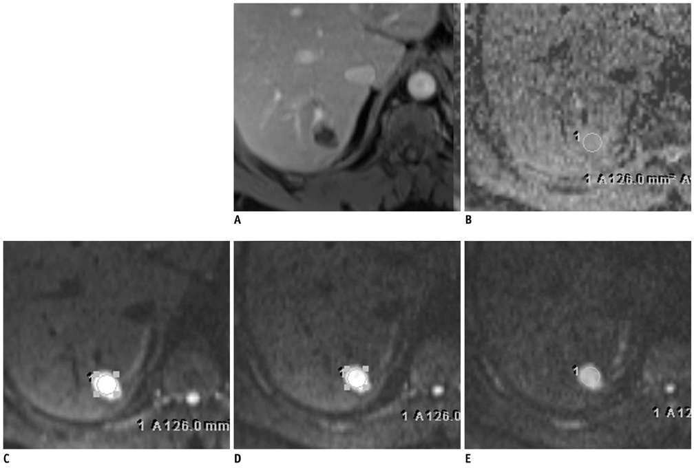
Fig. 2 Fifty four-year-old male patient with slowly enhancing hepatic hemangioma (group 3) (on S7, 21 mm in diameter). A. In portal phase of dynamic MRI, less than 25% contrast filling is seen, categorized as group 3. B. On ADC map, ADC value is automatically measured as 1.498 × 10⁻³ mm2/s. C-E. Based on signal intensities of DWI images with multiple b values (b = 50, 400, 800 s/mm2, respectively), mADC50-400 (1.134 × 10⁻³ mm2/s) is not higher than mADC400-800 (1.636 × 10⁻³ mm2/s). ADC = apparent diffusion coefficient, DWI = diffusion weighted imaging

Fig. 3 Box-plots of apparent diffusion coefficients (ADCs) measured in hepatic hemangiomas showing different speed (rapid, intermediate, or slow) of contrast enhancement during dynamic imaging. Decimal numbers on y-axis are mean ADCs (× 10-3 mm2/s) measured using region-of-interest on automatically-calculated ADC maps. Ranges of ADC values are overlapping each other in spite of significant difference of mean values between rapid and other lesions.

Fig. 4 Logarithmic plot of signal intensity against b value in each group (group 1, rapid; group 2, intermediate; group 3, slow). Slope of plot is represented by mADC50-400 (50-400 s/mm2) and mADC400-800 (400-800 s/mm2). mADC50-400 is higher, represented as stiffer slope, than mADC400-800 only in group 1 and results in slight bending b = 400 mm2/s which was not observed in other groups of hemangiomas showing straighter slope regardless of sizes of b factors. ADC = apparent diffusion coefficient.
Reference
-
1. Jeong MG, Yu JS, Kim KW. Hepatic cavernous hemangioma: temporal peritumoral enhancement during multiphase dynamic MR imaging. Radiology. 2000. 216:692–697.2. Vilgrain V, Boulos L, Vullierme MP, Denys A, Terris B, Menu Y. Imaging of atypical hemangiomas of the liver with pathologic correlation. Radiographics. 2000. 20:379–397.3. Ichikawa T, Haradome H, Hachiya J, Nitatori T, Araki T. Diffusion-weighted MR imaging with a single-shot echoplanar sequence: detection and characterization of focal hepatic lesions. AJR Am J Roentgenol. 1998. 170:397–402.4. Müller MF, Prasad P, Siewert B, Nissenbaum MA, Raptopoulos V, Edelman RR. Abdominal diffusion mapping with use of a whole-body echo-planar system. Radiology. 1994. 190:475–478.5. Namimoto T, Yamashita Y, Sumi S, Tang Y, Takahashi M. Focal liver masses: characterization with diffusion-weighted echo-planar MR imaging. Radiology. 1997. 204:739–744.6. Kim T, Murakami T, Takahashi S, Hori M, Tsuda K, Nakamura H. Diffusion-weighted single-shot echoplanar MR imaging for liver disease. AJR Am J Roentgenol. 1999. 173:393–398.7. Taouli B, Vilgrain V, Dumont E, Daire JL, Fan B, Menu Y. Evaluation of liver diffusion isotropy and characterization of focal hepatic lesions with two single-shot echo-planar MR imaging sequences: prospective study in 66 patients. Radiology. 2003. 226:71–78.8. Bruegel M, Holzapfel K, Gaa J, Woertler K, Waldt S, Kiefer B, et al. Characterization of focal liver lesions by ADC measurements using a respiratory triggered diffusion-weighted single-shot echo-planar MR imaging technique. Eur Radiol. 2008. 18:477–485.9. Gourtsoyianni S, Papanikolaou N, Yarmenitis S, Maris T, Karantanas A, Gourtsoyiannis N. Respiratory gated diffusion-weighted imaging of the liver: value of apparent diffusion coefficient measurements in the differentiation between most commonly encountered benign and malignant focal liver lesions. Eur Radiol. 2008. 18:486–492.10. Goshima S, Kanematsu M, Kondo H, Yokoyama R, Kajita K, Tsuge Y, et al. Hepatic hemangioma: correlation of enhancement types with diffusion-weighted MR findings and apparent diffusion coefficients. Eur J Radiol. 2009. 70:325–330.11. Yamada I, Aung W, Himeno Y, Nakagawa T, Shibuya H. Diffusion coefficients in abdominal organs and hepatic lesions: evaluation with intravoxel incoherent motion echo-planar MR imaging. Radiology. 1999. 210:617–623.12. Le Bihan D, Breton E, Lallemand D, Aubin ML, Vignaud J, Laval-Jeantet M. Separation of diffusion and perfusion in intravoxel incoherent motion MR imaging. Radiology. 1988. 168:497–505.13. Le Bihan D. Diffusion/perfusion MR imaging of the brain: from structure to function. Radiology. 1990. 177:328–329.14. Moteki T, Horikoshi H, Oya N, Aoki J, Endo K. Evaluation of hepatic lesions and hepatic parenchyma using diffusion-weighted reordered turboFLASH magnetic resonance images. J Magn Reson Imaging. 2002. 15:564–572.15. Yamashita Y, Ogata I, Urata J, Takahashi M. Cavernous hemangioma of the liver: pathologic correlation with dynamic CT findings. Radiology. 1997. 203:121–125.16. Obata S, Matsunaga N, Hayashi K, Ohtsubo M, Morikawa T, Takahara O. Fluid-fluid levels in giant cavernous hemangioma of the liver: CT and MRI demonstration. Abdom Imaging. 1998. 23:600–602.17. Ghai S, Dill-Macky M, Wilson S, Haider M. Fluid-fluid levels in cavernous hemangiomas of the liver: baffled? AJR Am J Roentgenol. 2005. 184:3 Suppl. S82–S85.18. Lee J, Lim HK, Jeon YH. Multiple hepatic hemangiomas with fluid-fluid levels. Australas Radiol. 2007. 51:Suppl. B310–B312.19. Soyer P, Bluemke DA, Fishman EK, Rymer R. Fluid-fluid levels within focal hepatic lesions: imaging appearance and etiology. Abdom Imaging. 1998. 23:161–165.20. Yu JS, Kim MJ, Kim KW. Intratumoral blood flow in cavernous hemangioma of the liver: radiologic-pathologic correlation. Radiology. 1998. 208:549–550.21. Kwee TC, Takahara T, Koh DM, Nievelstein RA, Luijten PR. Comparison and reproducibility of ADC measurements in breathhold, respiratory triggered, and free-breathing diffusion-weighted MR imaging of the liver. J Magn Reson Imaging. 2008. 28:1141–1148.22. Nasu K, Kuroki Y, Fujii H, Minami M. Hepatic pseudo-anisotropy: a specific artifact in hepatic diffusion-weighted images obtained with respiratory triggering. MAGMA. 2007. 20:205–211.
- Full Text Links
-
- Actions
-
Cited
- CITED
-
- Close
- Share
-
- Similar articles
-
- Correlation of the Speed of Enhancement of Hepatic Hemangiomas with Intravoxel Incoherent Motion MR Imaging
- Reversal of a Large Ischemic Lesion with Low Apparent Diffusion Coefficient Value by Rapid Spontaneous Recanalization
- Diffusion-weighted Imaging and Apparent Diffusion Coefficient Maps for the Evaluation of Pyogenic Ventriculitis
- Dysarthria with Hypoglycemia and Reversible Focal Hyperintensity Lesion on Diffusion?Weighted MRI
- Diffusion-weighted and Dynamic Contrast-enhanced MRI of Metastatic Bone Tumors: Correlation of the Apparent Diffusion Coefficient, K(trans) and ve values
