A Case of Hepatic Peribiliary Cysts in a Patient with Alcoholic Liver Cirrhosis
- Affiliations
-
- 1Department of Internal Medicine, Sanggye Paik Hospital, Inje University College of Medicine, Seoul, Korea. osbbang@paik.ac.kr
- KMID: 1387341
- DOI: http://doi.org/10.4166/kjg.2012.60.2.119
Abstract
- Hepatic peribiliary cysts (HPCs) are characterized by cystic dilatations of the peribiliary glands located throughout the branches of the biliary systems. Specifically, they are mainly located along the hepatic hilum and major portal tracts. The natural history and prognosis of HPCs are uncertain. In fact, almost all HPCs have been discovered incidentally during radiological examination or autopsy, and they are considered to be clinically harmless. Recently, several cases of HPCs associated with obstructive jaundice or liver failure were reported in patients with pre-existing liver disease in several studies. However, until now there have been no reports of such a case in Korea. Herein, we report a case of HPCs that show a disease course with a poor prognosis. These HPCs developed in a 47-year-old man with progressive alcoholic liver cirrhosis.
Keyword
MeSH Terms
Figure
-
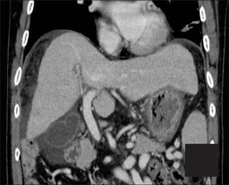
Fig. 1 Coronary view of abdominal computed tomography showed cirrhotic liver and ascites without hepatic peribiliary cysts.
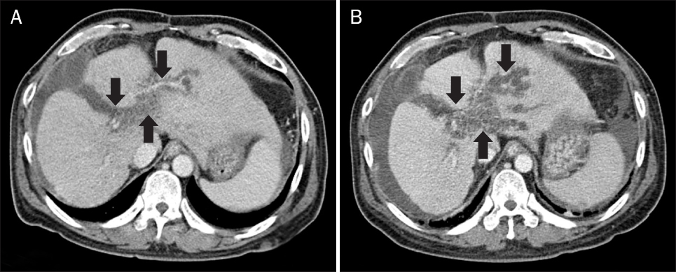
Fig. 2 Serial changes of follow-up abdominal computed tomography imaging. (A) Multiple, variable sized cystic lesions in the hepatic hilum and the large portal tracts (black arrows) were newly noted at the time of 38 months after diagnosis of alcoholic liver cirrhosis. (B) Hepatic peribiliary cysts (black arrows) increased in number and size at the time of 42 months after diagnosis of alcoholic liver cirrhosis.

Fig. 3 Magnetic resonance cholangiopancreaticography showed that multiple peribiliary cysts along the hepatic ducts, especially at the hepatic hilum and the left branch, represented as hyperintense on T2-weighted image. (A) T2-weighted coronary view (arrow). (B) T2-weighted axial view (arrow). (C) Maximum-intensity-projection reformation images.
Reference
-
1. Nakanuma Y, Kurumaya H, Ohta G. Multiple cysts in the hepatic hilum and their pathogenesis. A suggestion of periductal gland origin. Virchows Arch A Pathol Anat Histopathol. 1984. 404:341–350.2. Terada T, Nakanuma Y. Pathological observations of intrahepatic peribiliary glands in 1,000 consecutive autopsy livers. III. Survey of necroinflammation and cystic dilatation. Hepatology. 1990. 12:1229–1233.3. Kudo M. Hepatic peribiliary cysts: clinically harmless disease with potential risk due to gradual increase in size and number. J Gastroenterol. 2001. 36:286–288.4. Ikenaga N, Chijiiwa K, Otani K, Ohuchida J, Uchiyama S. A case of peribiliary cyst presenting with obstructive jaundice. J Gastrointest Surg. 2009. 13:174–176.5. Kai K, Eguchi Y, Kumagai T, Sugita Y, Tokunaga O. An autopsy case of obstructive jaundice due to hepatic multiple peribiliary cysts accompanying hepatolithiasis. Hepatol Res. 2008. 38:211–216.6. Johnson MA, Ravichandran P, Surendran R. Solitary extra-hepatic hilar peribiliary cyst presenting with obstructive jaundice: a case report. Acta Chir Belg. 2007. 107:716–719.7. Kim PJ, Kang DH, Jeong WJ, et al. A case of peribiliary cysts. Korean J Gastrointest Endosc. 2009. 38:368–370.8. Terada T, Minato H, Nakanuma Y, Shinozaki K, Kobayashi S, Matsui O. Ultrasound visualization of hepatic peribiliary cysts: a comparison with morphology. Am J Gastroenterol. 1992. 87:1499–1502.9. Nakanuma Y, Katayanagi K, Terada T, Saito K. Intrahepatic peribiliary glands of humans. I. Anatomy, development and presumed functions. J Gastroenterol Hepatol. 1994. 9:75–79.10. Hoshiba K, Matsui O, Kadoya M, et al. Peribiliary cysts in cirrhotic liver: observation on computed tomography. Abdom Imaging. 1996. 21:228–232.11. Itai Y, Ebihara R, Tohno E, et al. Hepatic peribiliary cysts: multiple tiny cysts within the larger portal tract, hepatic hilum, or both. Radiology. 1994. 191:107–110.12. Fujioka Y, Kawamura N, Tanaka S, Fujita M, Suzuki H, Nagashima K. Multiple hilar cysts of the liver in patients with alcoholic cirrhosis: report of three cases. J Gastroenterol Hepatol. 1997. 12:137–143.13. Seguchi T, Akiyama Y, Itoh H, et al. Multiple hepatic peribiliary cysts with cirrhosis. J Gastroenterol. 2004. 39:384–390.
- Full Text Links
-
- Actions
-
Cited
- CITED
-
- Close
- Share
-
- Similar articles
-
- Peribiliary cysts developed in normal underlying liver: report of a case
- A Case of Peribiliary Cysts
- Morphological Changes of Hepatic Microcirculation in N-diethylnitrosamine Induced Cirrhotic Rat Liver
- Peribiliary Cysts with Intrahepatic Bile Duct Obstruction: A Case Report
- Comparison of bacterial infection rate between patients with alcoholic liver cirrhosis and viral liver cirrhosis
