Korean J Radiol.
2008 Oct;9(5):391-395. 10.3348/kjr.2008.9.5.391.
Measuring Fractional Anisotropy of the Corpus Callosum Using Diffusion Tensor Imaging: Mid-Sagittal versus Axial Imaging Planes
- Affiliations
-
- 1Department of Radiology and the Research Institute of Radiological Science, Yonsei University College of Medicine, Seoul, Korea. eungykim@yuhs.ac
- 2Brain Korea 21 Project for Medical Science, Yonsei University College of Medicine, Seoul, Korea.
- 3School of Electrical and Electronic Engineering, Yonsei University, Seoul, Korea.
- KMID: 1385395
- DOI: http://doi.org/10.3348/kjr.2008.9.5.391
Abstract
OBJECTIVE
Many diffusion tensor imaging (DTI) studies of the corpus callosum (CC) have been performed with a relatively thick slice thickness in the axial plane, which may result in underestimating the fractional anisotropy (FA) of the CC due to a partial volume effect. We hypothesized that the FA of the CC can be more accurately measured by using mid-sagittal DTI. We compared the FA values of the CC between the axial and mid-sagittal DTI. MATERIALS AND METHODS: Fourteen healthy volunteers underwent MRI at 3.0 T. DTI was performed in both the mid-sagittal and axial planes. One 5-mm mid-sagittal image and twenty-five 2-mm axial images were obtained for the CC. The five regions of interest (ROIs) that included the prefrontal (I), premotor and supplementary motor (II), motor (III), sensory (IV) and parietal, temporal and occipital regions (V) were drawn along the border of the CC on each sagittal FA map. The FA values obtained from each region were compared between the two sagittal maps. RESULTS: The FA values of all the regions, except for region V, were significantly increased on the mid-sagittal imaging. The FA values in region IV were significantly underestimated on the mid-sagittal image from the axial imaging, compared with those in the regions I and V (p = 0.037 and p = 0.001, respectively). CONCLUSION: The FA values of the CC were significantly higher on the mid-sagittal DTI than those on the axial DTI in regions I-IV, and particularly in the region IV. Mid-sagittal DTI may provide more accurate FA values of the CC than can the axial DTI, and mid-sagittal DTI may be more desirable for studies that compare between patients and healthy subjects.
MeSH Terms
Figure
-
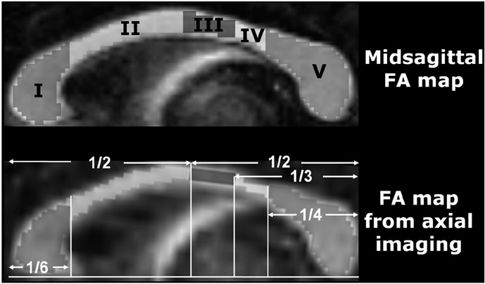
Fig. 1 Five regions of interest. Prefrontal (I), premotor and supplementary motor (II), motor (III), sensory (IV) and parietal, temporal and occipital regions (V) on mid-sagittal fractional anisotropy (FA) map (A) and the fractional anisotropy map from axial imaging (B).
Reference
-
1. Narr KL, Cannon TD, Woods RP, Thompson PM, Kim S, Asunction D, et al. Genetic contributions to altered callosal morphology in schizophrenia. J Neurosci. 2002. 22:3720–3729.2. Teipel SJ, Schapiro MB, Alexander GE, Krasuski JS, Horwitz B, Hoehne C, et al. Relation of corpus callosum and hippocampal size to age in nondemented adults with Down's syndrome . Am J Psychiatry. 2003. 160:1870–1878.3. Thompson PM, Dutton RA, Hayashi KM, Lu A, Lee SE, Lee JY, et al. 3D mapping of ventricular and corpus callosum abnormalities in HIV/AIDS. Neuroimage. 2006. 31:12–23.4. Filippi CG, Lin DD, Tsiouris AJ, Watts R, Packard AM, Heier LA, et al. Diffusion-tensor MR imaging in children with developmental delay: preliminary findings. Radiology. 2003. 229:44–50.5. Bozzali M, Falini A, Cercignani M, Baglio F, Farina E, Alberoni M, et al. Brain tissue damage in dementia with Lewy bodies: an in vivo diffusion tensor MRI study. Brain. 2005. 128:1595–1604.6. Thomas B, Eyssen M, Peeters R, Molenaers G, Van Hecke P, De Cock P, et al. Quantitative diffusion tensor imaging in cerebral palsy due to periventricular white matter injury. Brain. 2005. 128:2562–2577.7. Xie S, Xiao JX, Gong GL, Zang YF, Wang YH, Wu HK, et al. Voxel-based detection of white matter abnormalities in mild Alzheimer disease. Neurology. 2006. 66:1845–1849.8. Gupta RK, Saksena S, Hasan KM, Agarwal A, Haris M, Pandey CM, et al. Focal Wallerian degeneration of the corpus callosum in large middle cerebral artery stroke: serial diffusion tensor imaging. J Magn Reson Imaging. 2006. 24:549–555.9. Griffin CM, Chard DT, Ciccarelli O, Kapoor B, Barker GJ, Thompson AI, et al. Diffusion tensor imaging in early relapsingremitting multiple sclerosis. Mult Scler. 2001. 7:290–297.10. Ciccarelli O, Werring DJ, Barker GJ, Griffin CM, Wheeler-Kingshott CA, Miller DH, et al. A study of the mechanisms of normal-appearing white matter damage in multiple sclerosis using diffusion tensor imaging--evidence of Wallerian degeneration. J Neurol. 2003. 250:287–292.11. Pfefferbaum A, Rosenbloom MJ, Rohlfing T, Adalsteinsson E, Kemper CA, Deresinski S, et al. Contribution of alcoholism to brain dysmorphology in HIV infection: effects on the ventricles and corpus callosum. Neuroimage. 2006. 33:239–251.12. Pfefferbaum A, Adalsteinsson E, Sullivan EV. Dysmorphology and microstructural degradation of the corpus callosum: interaction of age and alcoholism. Neurobiol Aging. 2006. 27:994–1009.13. Pfefferbaum A, Rosenbloom MJ, Adalsteinsson E, Sullivan EV. Diffusion tensor imaging with quantitative fibre tracking in HIV infection and alcoholism comorbidity: synergistic white matter damage. Brain. 2007. 130:48–64.14. Sullivan EV, Adalsteinsson E, Pfefferbaum A. Selective agerelated degradation of anterior callosal fiber bundles quantified in vivo with fiber tracking. Cereb Cortex. 2006. 16:1030–1039.15. Hauser P, Dauphinais ID, Berrettini W, DeLisi LE, Gelernter J, Post RM. Corpus callosum dimensions measured by magnetic resonance imaging in bipolar affective disorder and schizophrenia. Biol Psychiatry. 1989. 26:659–668.16. Bastin ME, Armitage PA, Marshall I. A theoretical study of the effect of experimental noise on the measurement of anisotropy in diffusion imaging. Magn Reson Imaging. 1998. 16:773–785.17. Hofer S, Frahm J. Topography of the human corpus callosum revisited--comprehensive fiber tractography using diffusion tensor magnetic resonance imaging. Neuroimage. 2006. 32:989–994.18. Aboitiz F, Scheibel AB, Fisher RS, Zaidel E. Individual differences in brain asymmetries and fiber composition in the human corpus callosum. Brain Res. 1992. 598:154–161.19. Pierpaoli C, Basser PJ. Toward a quantitative assessment of diffusion anisotropy. Magn Reson Med. 1996. 36:893–906.20. Shimony JS, McKinstry RC, Akbudak E, Aronovitz JA, Snyder AZ, Lori NF, et al. Quantitative diffusion-tensor anisotropy brain MR imaging: normative human data and anatomic analysis. Radiology. 1999. 212:770–784.21. Oouchi H, Yamada K, Sakai K, Kizu O, Kubota T, Ito H, et al. Diffusion anisotropy measurement of brain white matter is affected by voxel size: underestimation occurs in areas with crossing fibers. AJNR Am J Neuroradiol. 2007. 28:1102–1106.
- Full Text Links
-
- Actions
-
Cited
- CITED
-
- Close
- Share
-
- Similar articles
-
- Measurement of Fractional Anisotropy in Normal Cerebral White Matter and Brain Tumors with Diffusion Tensor Imaging
- Assessment of Normal-Appearing White Matter Damage in Multiple Sclerosis Using Diffusion Tensor Imaging
- The Effect of Imaging Parameters of Diffusion Tensor Imaging on Fractional Anisotropy
- Anisotropy Measurement and Fiber Tracking of the White Matter by Using Diffusion Tensor MR Imaging: Influence of the Number of Diffusion-Sensitizing Gradient Direction
- The Contemplation of Neuropathological Abnormalities of the Corpus Callosum in Schizophrenia: A Diffusion Tensor Imaging Study
