Intracardiac Eustachian Valve Cyst in an Adult Detected with Other Cardiac Anomalies: Usefulness of Multidetector CT in Diagnosis
- Affiliations
-
- 1Department of Radiology, Seoul St. Mary's Hospital, College of Medicine, The Catholic University of Korea, Seoul 137-701, Korea. jijung@catholic.ac.kr
- 2Department of Thoracic and Cardiovascular Surgery, Seoul St. Mary's Hospital, College of Medicine, The Catholic University of Korea, Seoul 137-701, Korea.
- 3Department of Pathology, Seoul St. Mary's Hospital, College of Medicine, The Catholic University of Korea, Seoul 137-701, Korea.
- KMID: 1383864
- DOI: http://doi.org/10.3348/kjr.2012.13.4.500
Abstract
- We present an unusual case of an intracardiac Eustachian valve cyst observed concurrently with atresia of the coronary sinus ostium, a persistent left superior vena cava (LSVC) and a bicuspid aortic valve. There have been several echocardiographic reports of Eustachian valve cysts; however, there is no report of multidetector computed tomography (MDCT) findings related to a Eustachian valve cyst. Recently, we observed a Eustachian valve cyst diagnosed on MDCT showing a hypodense cyst at the characteristic location of the Eustachian valve (the junction of the right atrium and inferior vena cava). MDCT also demonstrated additional cardiovascular anomalies including atresia of the coronary sinus ostium and a persistent LSVC and bicuspid aortic valve.
Keyword
MeSH Terms
-
Aged
Aortic Valve/abnormalities/radiography
Cysts/*radiography
Echocardiography, Transesophageal
Heart Atria/abnormalities/radiography
Heart Defects, Congenital/*radiography/surgery
Humans
Male
*Tomography, X-Ray Computed
Vena Cava, Inferior/abnormalities/radiography
Vena Cava, Superior/abnormalities/radiography
Figure
-
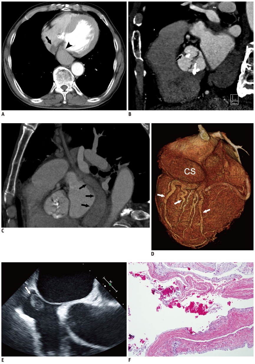
Fig. 1 Intracardiac Eustachian valve cyst in 71-year-old man with other cardiac anomaly including atresia of coronary sinus ostium, persistent left superior vena cava and bicuspid aortic valve. A. Axial contrast-enhanced chest CT shows 1.7-cm lobulating contoured hypodense mass at junction of right atrium and inferior vena cava (arrow). Note dilated coronary sinus (arrowhead). B. Cardiac CT with multiplanar reformatted image shows bicuspid aortic valve with raphe (arrow) and dense calcification, suggesting aortic stenosis. C. Sagittal multiplanar reformatted image of cardiac CT shows dilated coronary sinus connected to persistent left superior vena cava (arrows), which drains into left innominate vein (not shown). D. Volume-rendered CT image shows dilated coronary sinus (CS) and cardiac veins (arrows). E. Transesophageal echocardiogram shows 2 × 1-cm oval echogenic mass attached to right atrium wall that contains internal anechoic cystic lesion (arrow). F. Pathology shows (H & E stain × 100) pseudocyst induced through fibromyxoid degeneration and flattening of wall with blood.
Reference
-
1. Yater WM. Variations and anomalies of the venous valves of the right atrium of the human heart. Arch Pathol. 1929. 7:418–441.2. Watson T, Kakar P, Srivastava S, Dhanjal TS. Eustachian valve remnant. Cardiol J. 2007. 14:508–509.3. Cujec B, Ulmer B, McKaigney JP, Bharadwaj B. Right atrial myxoma presenting as Budd-Chiari syndrome. Ann Thorac Surg. 1987. 44:658–659.4. Nkomo VT, Miller FA. Eustachian valve cyst. J Am Soc Echocardiogr. 2001. 14:1224–1226.5. Gilkeson RC, Ciancibello L, Zahka K. Pictorial essay. Multidetector CT evaluation of congenital heart disease in pediatric and adult patients. AJR Am J Roentgenol. 2003. 180:973–998.6. Otsuka K, Terasaki F, Iimori A, Tonari S, Shimomura H, Ito T, et al. Right atrial blood cyst with total occlusion of the right coronary artery. Heart Vessels. 2007. 22:208–210.7. Boyd TA. Blood cysts on the heart valves of infants. Am J Pathol. 1949. 25:757–759.8. Sakakibara S, Katsuhara K, Iida Y, Nishida H. Pulmonary subvalvular tumor. Dis Chest. 1967. 51:637–642.9. Scheffel H, Baumueller S, Stolzmann P, Leschka S, Plass A, Alkadhi H, et al. Atrial myxomas and thrombi: comparison of imaging features on CT. AJR Am J Roentgenol. 2009. 192:639–645.10. Nakamura M, Urita R, Okamoto F, Abe T, Komatsu S. [A case of right atrial myxoma, originating from the eustachian valve]. Kyobu Geka. 1990. 43:920–923.11. Grebenc ML, Rosado-de-Christenson ML, Green CE, Burke AP, Galvin JR. Cardiac myxoma: imaging features in 83 patients. Radiographics. 2002. 22:673–689.12. Loukas M, Sullivan A, Tubbs RS, Weinhaus AJ, Derderian T, Hanna M. Chiari's network: review of the literature. Surg Radiol Anat. 2010. 32:895–901.13. Jha NK, Gogna A, Tan TH, Wong KY, Shankar S. Atresia of coronary sinus ostium with retrograde drainage via persistent left superior vena cava. Ann Thorac Surg. 2003. 76:2091–2092.14. Santoscoy R, Walters HL 3rd, Ross RD, Lyons JM, Hakimi M. Coronary sinus ostial atresia with persistent left superior vena cava. Ann Thorac Surg. 1996. 61:879–882.
- Full Text Links
-
- Actions
-
Cited
- CITED
-
- Close
- Share
-
- Similar articles
-
- Coronary Artery Anomaly, What Radiologist Should Know?
- Aortic Stenosis: Evaluation with Multidetector CT Angiography and MR Imaging
- A Case of Intracardiac Metastasis of Hepatocellular Carcinoma Presenting with Functional Tricuspid Valve Stenosis Accompanied with Hepatopulmonary Syndrome
- Blood Cyst of Subvalvular Apparatus of the Mitral Valve in an Adult
- Aortic Arch Variants and Anomalies: Embryology, Imaging Findings, and Clinical Considerations
