Eosinophilic Otitis Media: CT and MRI Findings and Literature Review
- Affiliations
-
- 1Department of Radiology and Research Institute of Radiology, University of Ulsan College of Medicine, Asan Medical Center, Seoul 138-736, Korea. jeonghlee@amc.seoul.kr
- 2Department of Otorhinolaryngology, University of Ulsan College of Medicine, Asan Medical Center, Seoul 138-736, Korea.
- 3Department of Pathology, University of Ulsan College of Medicine, Asan Medical Center, Seoul 138-736, Korea.
- KMID: 1372859
- DOI: http://doi.org/10.3348/kjr.2012.13.3.363
Abstract
- Eosinophilic otitis media (EOM) is a relatively rare, intractable, middle ear disease with extremely viscous mucoid effusion containing eosinophils. EOM is associated with adult bronchial asthma and nasal allergies. Conventional treatments for otitis media with effusion (OME) or for chronic otitis media (COM), like tympanoplasty or mastoidectomy, when performed for the treatment of EOM, can induce severe complications such as deafness. Therefore, it should be differentiated from the usual type of OME or COM. To our knowledge, the clinical and imaging findings of EOM of temporal bone are not well-known to radiologists. We report here the CT and MRI findings of two EOM cases and review the clinical and histopathologic findings of this recently described disease entity.
MeSH Terms
Figure
-
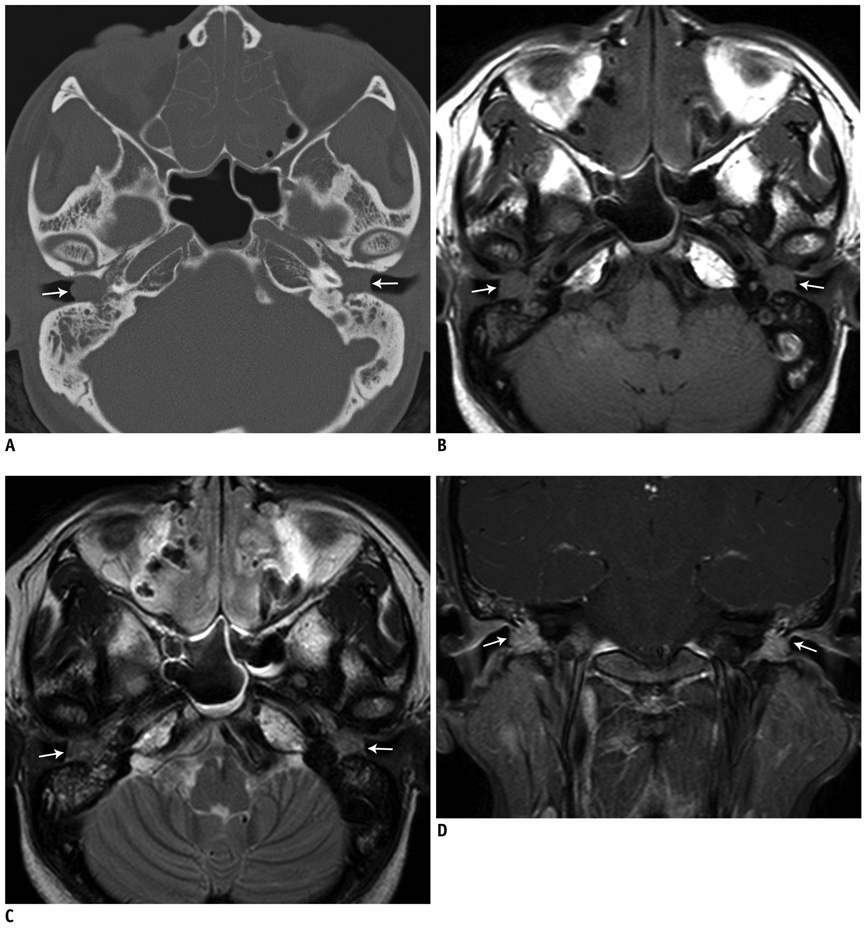
Fig. 1 37-year-old woman with progressive, bilateral hearing disturbance. (A) TBCT shows complete soft-tissue filling in bilateral middle ear cavities (arrow) and sclerotic mastoid. No bony erosion, except for minimal marginal erosion of ossicular chain is seen. Ethmoid sinuses are completely obliterated by thickened mucosa, which is also seen in sphenoid sinuses. (B-D) TBMR shows T1 low- (B) and T2 intermediate- (C) signal-intensity soft-tissue lesions filling both middle ear cavities (arrow) with mild heterogeneous contrast enhancement (D) suggesting granulation tissue. TBCT = temporal bone CT

Fig. 2 33-year-old man with persistent otorrhea. A, B. Axial and coronal TBCT images demonstrate soft-tissue lesions bulging from bilateral middle-ear cavities and filling mastoid air cells. There is no evidence of bony erosion except for bony defect at right mastoid from previous surgery. Mucosal thickening of bilateral maxillary sinus is also shown. C, D. Bilateral Eustachian tubes are clearly traced on serial coronal TBCT images (arrows). TBCT = temporal bone CT
Reference
-
1. Sipilä P, Karma P. Inflammatory cells in mucoid effusion of secretory otitis media. Acta Otolaryngol. 1982. 94:467–472.2. Iino Y, Usubuchi H, Kodama K, Kanazawa H, Takizawa K, Kanazawa T, et al. Eosinophilic inflammation in the middle ear induces deterioration of bone-conduction hearing level in patients with eosinophilic otitis media. Otol Neurotol. 2010. 31:100–104.3. Iino Y, Kakizaki K, Saruya S, Katano H, Komiya T, Kodera K, et al. Eustachian tube function in patients with eosinophilic otitis media associated with bronchial asthma evaluated by sonotubometry. Arch Otolaryngol Head Neck Surg. 2006. 132:1109–1114.4. Iwasaki S, Nagura M, Mizuta K. Cochlear implantation in a patient with eosinophilic otitis media. Eur Arch Otorhinolaryngol. 2006. 263:365–369.5. Nagamine H, Iino Y, Kojima C, Miyazawa T, Iida T. Clinical characteristics of so called eosinophilic otitis media. Auris Nasus Larynx. 2002. 29:19–28.6. Iino Y, Tomioka-Matsutani S, Matsubara A, Nakagawa T, Nonaka M. Diagnostic criteria of eosinophilic otitis media, a newly recognized middle ear disease. Auris Nasus Larynx. 2011. 38:456–461.
- Full Text Links
-
- Actions
-
Cited
- CITED
-
- Close
- Share
-
- Similar articles
-
- A Case of Remission of Refractory Eosinophilic Otitis Media Treated With Dupilumab
- Development of Animal Models of Otitis Media
- Prognostic significance of mastoid pneumatization in childhood otitis media with effusion: temporal bone CT evaluation
- Eosinophilic Otitis Media 3 Cases Discovered with Recurrent and Sticky Otorrhea after Ventilation Tube Insertion
- Two Cases of Eosinophilic Otitis Media
