Treatment of a Recurrent Chest Wall Desmoid Tumor Using a CT-Guided Steroid Injection
- Affiliations
-
- 1Department of Radiology, Soonchunhyang University Bucheon Hospital, Bucheon 420-767, Korea. radpsh@schmc.ac.kr
- 2Department of Thoracic Surgery, Soonchunhyang University Bucheon Hospital, Bucheon 420-767, Korea.
- KMID: 1372854
- DOI: http://doi.org/10.3348/kjr.2012.13.3.342
Abstract
- We report on a 41-year-old woman with a chest wall desmoid tumour who was successfully treated with a computed tomography (CT)-guided steroid injection. She presented with a palpable mass in the right upper chest wall and was treated by surgical excision and postoperative radiation therapy due to recurrence of the mass at the surgical site. At 20 months after the second operation, a recurrent mass was again detected in the anterosuperior portion of the previous surgical site on CT. We performed a CT-guided steroid injection weekly for 4 weeks by applying a mixture of 3 mL of triamcinolone acetonide (40 mg/mL) and 3 mL of 1% Lidocaine, administering 4-6 mL of the mixture, to the lesion. Six months later, CT showed a marked decrease in the size of the mass.
Keyword
MeSH Terms
Figure
-
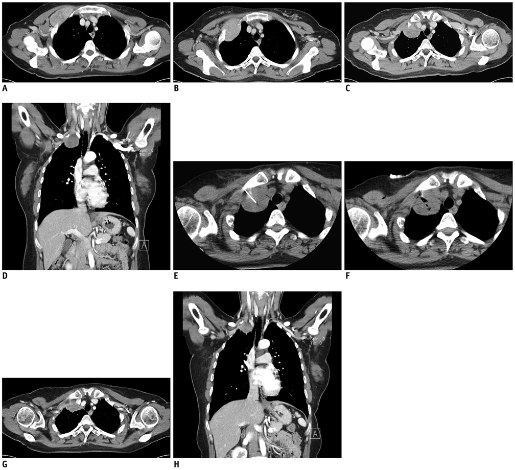
Fig. 1 Recurrent chest wall desmoid tumor in 41-year-old woman. A. Contrast enhanced axial CT image shows round, 2.4 × 6-cm enhancing mass surrounded by anterior arc of right second rib. B. Contrast enhanced axial CT image shows 6 × 4-cm homogeneously enhancing mass at previous surgical site with cortical disruption at anterior arc of right first and second ribs. C, D. Contrast enhanced axial CT and coronal reconstruction images show 3.5 × 3.2 × 3.2-cm recurrent mass in anterosuperior portion of previous surgical site with heterogeneous enhancement. E, F. Axial CT image shows injection needle located in center of mass and CT image after injection. G, H. Follow-up chest axial CT and coronal reconstruction images after 6 months show marked decrease in size of mass.
Reference
-
1. Kujak JL, Liu PT, Johnson GB, Callstrom MR. Early experience with percutaneous cryoablation of extra-abdominal desmoid tumors. Skeletal Radiol. 2010. 39:175–182.2. McDonald ES, Yi ES, Wenger DE. Best cases from the AFIP: extraabdominal desmoid-type fibromatosis. Radiographics. 2008. 28:901–906.3. Dinauer PA, Brixey CJ, Moncur JT, Fanburg-Smith JC, Murphey MD. Pathologic and MR imaging features of benign fibrous soft-tissue tumors in adults. Radiographics. 2007. 27:173–187.4. Nuyttens JJ, Rust PF, Thomas CR Jr, Turrisi AT 3rd. Surgery versus radiation therapy for patients with aggressive fibromatosis or desmoid tumors: a comparative review of 22 articles. Cancer. 2000. 88:1517–1523.5. Al-Attar A, Mess S, Thomassen JM, Kauffman CL, Davison SP. Keloid pathogenesis and treatment. Plast Reconstr Surg. 2006. 117:286–300.6. Sherris DA, Larrabee WF Jr, Murakami CS. Management of scar contractures, hypertrophic scars, and keloids. Otolaryngol Clin North Am. 1995. 28:1057–1068.
- Full Text Links
-
- Actions
-
Cited
- CITED
-
- Close
- Share
-
- Similar articles
-
- CT findings of Desmoid tumor arising at Abdominai Wall
- Intrathoracic Desmoid Tumor: A Case Report and Radiological Evaluation
- Desmoid Tumor of the Facet Joint: A Case Report
- Seven Cases of Desmoid Tumor of Trunk
- Intrathoracic Desmoid Tumor Presenting as Multiple Lung Nodules 13 Years after Previous Resection of Abdominal Wall Desmoid Tumor
