A Developmental Mechanism of Spontaneous Reattachment in Rhegmatogenous Retinal Detachment
- Affiliations
-
- 1Department of Ophthalmology, Samsung Medical Center, Sungkyunkwan University School of Medicine, Seoul, Korea. swkang@skku.edu
- KMID: 1364868
- DOI: http://doi.org/10.3341/kjo.2012.26.2.135
Abstract
- This retrospective observational case series on eyes from three patients was done to elucidate the developmental mechanism of spontaneous reattachment of rhegmatogenous retinal detachment (SRRRD). The study eyes of each patients showed evidence of retinal break and diffuse retinal pigmentary change. Ultrasound biomicroscopic examination revealed vitreous fibers attached to the area around the retinal break. Posterior vitreous attachment was confirmed in each eye. A thin fibrovascular membrane incompletely sealing the retinal break was noted in one case. We suggest that the vitreous attachment around the retinal break and the sealing of the break with adjacent vitreous fibers seem to be involved in the developmental mechanism of SRRRD.
MeSH Terms
-
Adult
Atrophy
Disease Progression
Female
Humans
Male
Middle Aged
Remission, Spontaneous
Retina/*abnormalities/pathology/*physiopathology
Retinal Detachment/*etiology/pathology/*physiopathology
Retinal Pigment Epithelium/abnormalities/pathology/physiopathology
Retrospective Studies
Vitreous Body/abnormalities/pathology/physiopathology
Young Adult
Figure
-
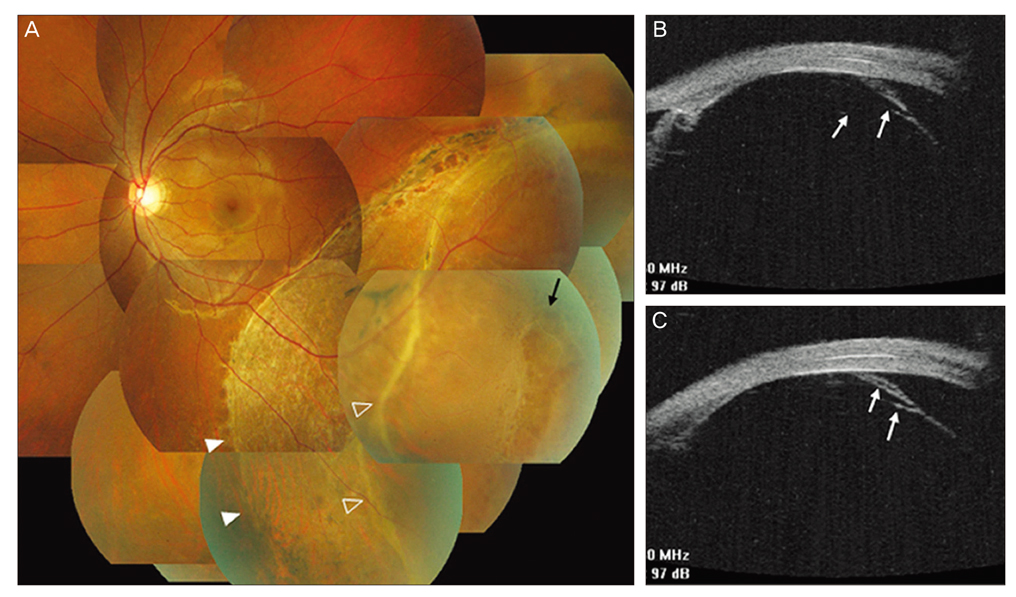
Fig. 1 Left eye in patient 1. (A) Fundus photograph at presentation: pigmentary clumping and atrophy at the attached retina (solid white arrow heads) posterior to the area of retinal detachment (empty arrow heads) with a retinal hole (black arrow). (B,C) Ultrasound biomicroscopic examination disclosed vitreous fibers (white arrows) both anterior and posterior to the retinal hole.
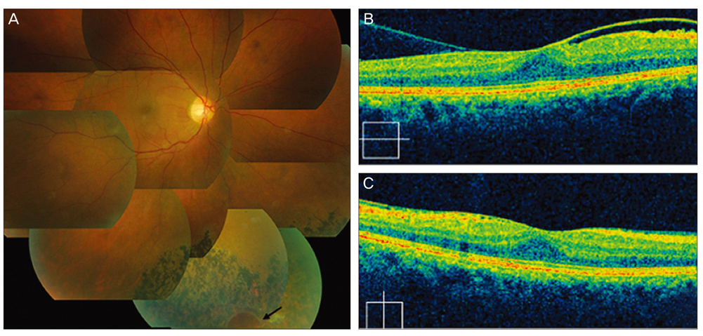
Fig. 2 Right eye in patient 2. (A) Notice the retinal pigmentary alteration and retinal hole (black arrow) at the 5-o'clock position. (B,C) Optical coherence tomographic examination revealed development of posterior vitreous detachment on the temporal side of the fovea only.
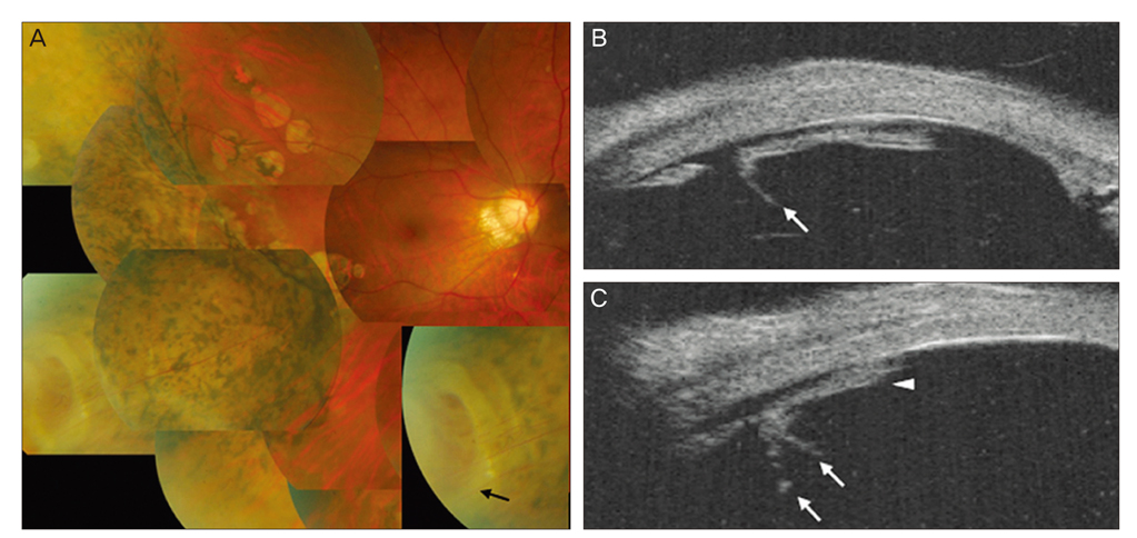
Fig. 3 Right eye in patient 3. (A) Fundus photograph at presentation shows extensive retinal pigmentary clumping, atrophy, and a 2 disc-diameter horseshoe retinal tear at the 8-o'clock position. The magnified image reveals thin fibrovascular membrane (black arrow) connecting the anterior and posterior margins of the retinal tear. (B,C) Ultrasound biomicroscopic imaging showed vitreous fibers (white arrows) and membranous structure sealing the retinal tear (white arrow head).
Cited by 1 articles
-
Two Cases of Acute Spontaneous Resolution in Macula-Off Rhegmatogenous Retinal Detachment
Jung Yul Park, Min Kyu Shin, Sung Who Park, Ik Soo Byon, Ji Eun Lee
J Korean Ophthalmol Soc. 2015;56(3):466-470. doi: 10.3341/jkos.2015.56.3.466.
Reference
-
1. Cantrill HL. Spontaneous retinal reattachment. Retina. 1981. 1:216–219.2. De Juan E Jr, Machemer R. Spontaneous reattachment of the retina despite proliferative vitreoretinopathy. Am J Ophthalmol. 1984. 97:428–433.3. Kokolakis SN, Bravo L, Chignell AH. Late retinal reattachment. Br J Ophthalmol. 1981. 65:142–146.4. Cho HY, Chung SE, Kim JI, et al. Spontaneous reattachment of rhegmatogenous retinal detachment. Ophthalmology. 2007. 114:581–586.5. Gonzales CR, Gupta A, Schwartz SD, Kreiger AE. The fellow eye of patients with phakic rhegmatogenous retinal detachment from atrophic holes of lattice degeneration without posterior vitreous detachment. Br J Ophthalmol. 2004. 88:1400–1402.
- Full Text Links
-
- Actions
-
Cited
- CITED
-
- Close
- Share
-
- Similar articles
-
- Pathology of Rhegmatogenous Retinal Detachment
- Spontaneous reattachment of retinal detachment with macular hole in nonmyopic patients
- Rhegmatogenous Retinal Detachment Associated with Atopic Dermatitis
- Clinical Evaluation of Choroidal Detachment Associated with Rhegmatogenous Retinal Detachment
- Factors Affecting the Visual Outcome after Successful Retinal Reattachment Surgery in Rhegmatogenous Retinal Detachment Involving the Macula
