Evaluation of the Sigmoid Notch Involvement in the Intra-Articular Distal Radius Fractures: The Efficacy of Computed Tomography Compared with Plain X-ray
- Affiliations
-
- 1Department of Orthopedic Surgery, Konyang University Hospital, Daejeon, Korea. oeo-oeoeo@hanmail.net
- KMID: 1245403
- DOI: http://doi.org/10.4055/cios.2012.4.1.83
Abstract
- BACKGROUND
The purpose of this study is to evaluate the efficacy of computed tomography (CT) scans compared with plain radiographs on detecting the involvement of the sigmoid notch.
METHODS
This study involved 121 cases diagnosed as the intra-articular distal radius fracture and performed post-reduction CT scans. We determined the presence of the sigmoid notch involvement with both plain radiographs and CT scans and compared findings of plain radiographs with CT scans about the incidence and the pattern of injuries. And the differences of results between arbeitsgemeinschaft fur osteosyntheses (AO) type C2 and C3 were compared.
RESULTS
The incidences of sigmoid notch involvement detected in plain radiographs were 81 cases (66.9%), whereas CT scans were 99 cases (81.9%). The sensitivity of plain radiographs compared with CT scans was 74.7%, the specificity was 68.2%, the positive predictive value was 91.4%, the negative predictive value was 37.5%, the false negative value was 25.3%, and the false positive value was 31.8%. In comparison between AO type C2 and C3, the incidence of sigmoid notch involvement was not a significant difference, but the displacement of fracture fragment showed a significant difference.
CONCLUSIONS
The intra-articular distal radius fracture usually accompanies the sigmoid notch involvement. Considering that the evaluation of sigmoid notch involvement by plain radiography often results in misinterpretation or underestimation, performing CT scan in intra-articular distal radius fracture is thought to be beneficial.
MeSH Terms
Figure
-
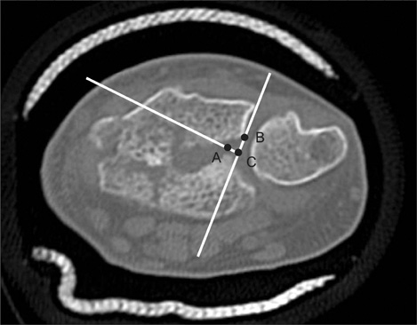
Fig. 1 Axial computed tomography image of the distal radius showing a displaced fracture of the sigmoid notch (SN). Point A and B were marked at subchondral fracture margins of SN. First line was drawn between the dorsal corner of SN and point B. Second line through point A was perpendicular to first line. The intersection of two lines was point C. Step-off of SN was measured as the distance between point A and C. Gap displacement was measured as the distance between point B and C.
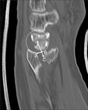
Fig. 2 Sagittal computed tomography image of the radiocarpal joint. Point A and C were marked at subchondral fracture margin of lunate facet of distal radius. A circle matching curvature of remaining articular surface of distal radius was traced and passed through point A. First line was drawn from center of circle to point C. The intersection between circle and first line was point B. Step displacement is measured as the distance between points B and C. Gap displacement is measured as the distance between points A and B.
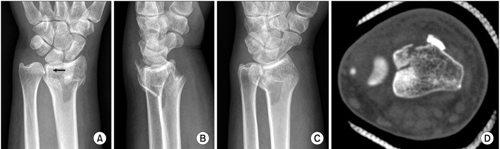
Fig. 3 Radiographs of 57-year-old woman with left distal radius fracture. Initial radiographs (A-C) were showing intra-articular fracture line (black arrow on image (A)) and metaphyseal comminution. But, authors misinterpreted the involvement of the sigmoid notch presented because of overlaps of ulnar head and lunate facet on lateral view (B) or dorsal-ulnar corner of distal radius on oblique view (C). The fracture of the sigmoid notch with displaced fragment was confirmed on axial computed tomography image (D).
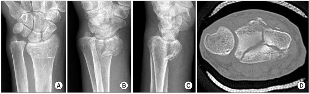
Fig. 4 Initial radiographs (A-C) of 79-year-old man were showing intra-articular fracture of left distal radius fracture. The authors interpreted the disruption of the sigmoid notch with displacement of fracture fragment on image A-C. But, the sigmoid notch was intact and intra-articular fracture line was located near the sigmoid notch on axial computed tomography image (D).
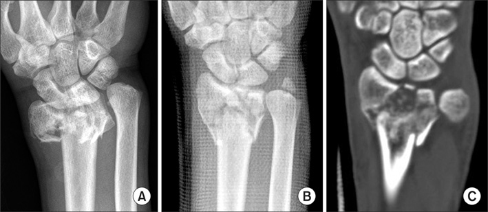
Fig. 5 Distal radius fracture involved the articular surface of radiocarpal and distal radioulnar joint. This case was classified as arbeitsgemeinschaft für osteosyntheses type C3 and Ronzental type 3a. Axial-plane fracture line of the sigmoid notch can be easily found in coronal computed tomography image (C) than pre-reduction (A) or post-reduction (B) radiographs.
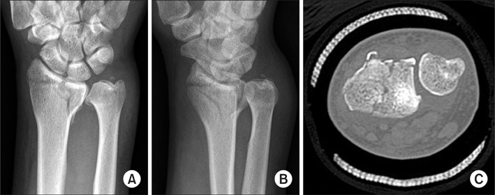
Fig. 6 Initial radiographs (A and B) and axial CT image (C) showing intra-articular distal radius fracture. This case was classified as arbeitsgemeinschaft für osteosyntheses type C2 and Rozental type 1b. Dorso-ulnar cortical corner fracture of the distal radius was not found on postero-anterior view (A) but oblique view (B). And minimal involvement of the sigmoid notch was confirmed on axial computed tomography image (C).
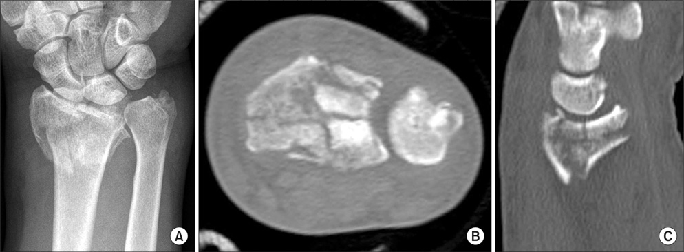
Fig. 7 Comminuted distal radius fracture was classified as arbeitsgemeinschaft für osteosyntheses type C3 and Rozental type 3b. An accurate assessment is difficult about sigmoid notch in plane radiograph (A). Axial (B) and sagittal (C) computed tomography image shows comminuted fracture of articular surface of the sigmoid notch.
Reference
-
1. Turner RG, Faber KJ, Athwal GS. Complications of distal radius fractures. Hand Clin. 2010. 26(1):85–96.
Article2. Medoff RJ. Essential radiographic evaluation for distal radius fractures. Hand Clin. 2005. 21(3):279–288.
Article3. Katz MA, Beredjiklian PK, Bozentka DJ, Steinberg DR. Computed tomography scanning of intra-articular distal radius fractures: does it influence treatment. J Hand Surg Am. 2001. 26(3):415–421.
Article4. Johnston GH, Friedman L, Kriegler JC. Computerized tomographic evaluation of acute distal radial fractures. J Hand Surg Am. 1992. 17(4):738–744.
Article5. Catalano LW 3rd, Barron OA, Glickel SZ. Assessment of articular displacement of distal radius fractures. Clin Orthop Relat Res. 2004. (423):79–84.
Article6. Cole RJ, Bindra RR, Evanoff BA, Gilula LA, Yamaguchi K, Gelberman RH. Radiographic evaluation of osseous displacement following intra-articular fractures of the distal radius: reliability of plain radiography versus computed tomography. J Hand Surg Am. 1997. 22(5):792–800.
Article7. Pruitt DL, Gilula LA, Manske PR, Vannier MW. Computed tomography scanning with image reconstruction in evaluation of distal radius fractures. J Hand Surg Am. 1994. 19(5):720–727.
Article8. Trumble TE, Schmitt SR, Vedder NB. Factors affecting functional outcome of displaced intra-articular distal radius fractures. J Hand Surg Am. 1994. 19(2):325–340.
Article9. Geissler WB, Fernandez DL, Lamey DM. Distal radioulnar joint injuries associated with fractures of the distal radius. Clin Orthop Relat Res. 1996. (327):135–146.
Article10. Rozental TD, Bozentka DJ, Katz MA, Steinberg DR, Beredjiklian PK. Evaluation of the sigmoid notch with computed tomography following intra-articular distal radius fracture. J Hand Surg Am. 2001. 26(2):244–251.
Article11. Shrout PE, Fleiss JL. Intraclass correlations: uses in assessing rater reliability. Psychol Bull. 1979. 86(2):420–428.
Article12. Knirk JL, Jupiter JB. Intra-articular fractures of the distal end of the radius in young adults. J Bone Joint Surg Am. 1986. 68(5):647–659.
Article13. Taras JS, Ladd AL, Kalainov DM, Ruch DS, Ring DC. New concepts in the treatment of distal radius fractures. Instr Course Lect. 2010. 59:313–332.14. Kihara H, Palmer AK, Werner FW, Short WH, Fortino MD. The effect of dorsally angulated distal radius fractures on distal radioulnar joint congruency and forearm rotation. J Hand Surg Am. 1996. 21(1):40–47.
Article15. Nagy L. Salvage of post-traumatic arthritis following distal radius fracture. Hand Clin. 2005. 21(3):489–498.
Article16. Frykman G. Fracture of the distal radius including sequelae--shoulder-hand-finger syndrome, disturbance in the distal radio-ulnar joint and impairment of nerve function: a clinical and experimental study. Acta Orthop Scand. 1967. Suppl 108. 3+.
Article17. Harreld KL, Apel P, Koman LA, Li Z. Lunate-lunate facet ratio and variance to predict articular gap after distal radius fracture. J Hand Surg Am. 2009. 34(9):1625–1632.
Article18. Lozano-Calderon SA, Doornberg J, Ring D. Fractures of the dorsal articular margin of the distal part of the radius with dorsal radiocarpal subluxation. J Bone Joint Surg Am. 2006. 88(7):1486–1493.
Article
- Full Text Links
-
- Actions
-
Cited
- CITED
-
- Close
- Share
-
- Similar articles
-
- The Usefulness of Computed Tomography in Distal Radius Fractures Involving Articular Surface
- Clinical Assessment of the Distal Radioulnar Joint Instability After Treatment of Intra-articular Fractures of the Distal Radius using Computed Tomography
- Treatment of the distal radius fractures with open reduction and internal fixation
- A Clinical Study of Anatomic Position Change on the Intra - articular Comminuted Fracture of the Distal Radius
- Intra-articular fracture reduction: a comparative observational study of clinical results after the surgical treatment of distal radius fractures
