Syringocystadenocarcinoma Papilliferum: A Case Report
- Affiliations
-
- 1Department of Dermatology, College of Medicine, Yeungnam University, Daegu, Korea. dhshin@med.yu.ac.kr
- KMID: 1127103
- DOI: http://doi.org/10.3346/jkms.2007.22.4.762
Abstract
- Syringocystadenocarcinoma papilliferum (SCACP) is a rare form of adenocarcinoma of the skin. This is the malignant counterpart of syringocystadenoma papilliferum (SCAP) and usually develops on the scalp in a long-standing lesion identified clinically as SCAP. We describe a 65-yr-old Korean man with a nodule on the right supra-pubic area with a 2-yr duration. Histologically this tumor had a similar overall configuration as in SCAP, but the tumor was asymmetric and poorly circumscribed, extending into the deep dermis and showed cytologic atypia. The tumor cells showed positive reaction to GCDFP-15, but negative reaction to CEA and HMFG-1. We established the diagnosis of SCACP in the patient, and a wide excision was performed to remove the tumor. The patient has been well without relapse or metastasis for 2 yr.
Keyword
MeSH Terms
Figure
-
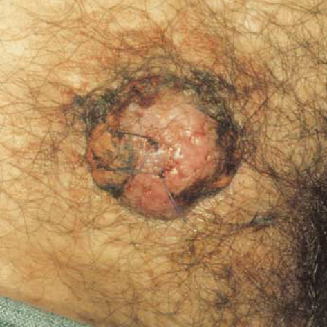
Fig. 1 A single 3.4×3.5 cm-sized erythematous dome-shaped nodule covered with bloody crust on the right suprapubic area.
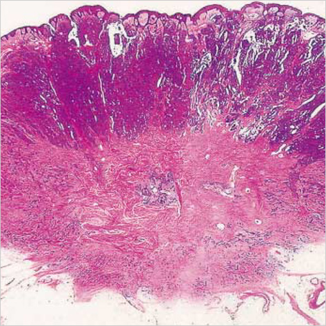
Fig. 2 A low-power view of the lesion shows a tumor with a solid and cystic pattern in the upper dermis and diffusely infiltrating the lower dermis (H&E, ×1).
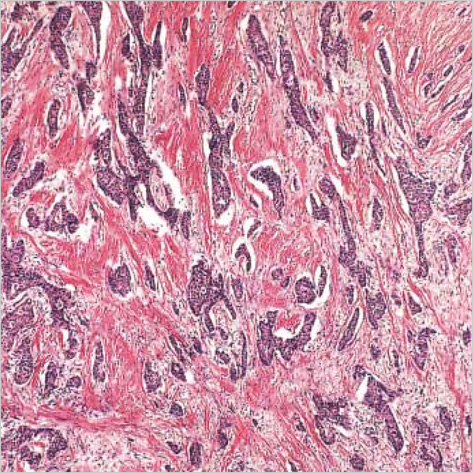
Fig. 3 Tumor cells of strand or cord structures in the deep reticular dermis with desmoplastic stroma (H&E, ×40).
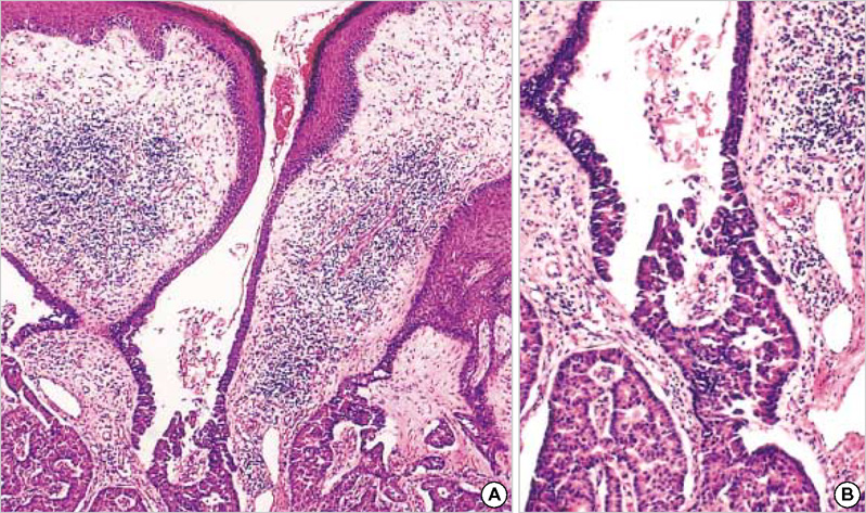
Fig. 4 (A) Cystic invagination connected to the infundibular portion extends downward (H&E, ×40). (B) Inferior portion of cystic invagination lined by multiple layered epithelium with decapitation on the luminal surface of the cells, numerous plasma cells and lymphocytes in the stroma (H&E, ×100).
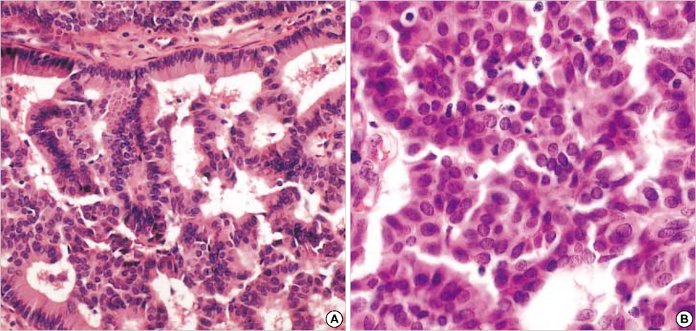
Fig. 5 (A) Tumor cells having large and hyperchromatic nuclei (H&E, ×200). (B) Hyperchromatic nuclei and mitotic figures were observed in these tumor cells (H&E, ×200).
Cited by 1 articles
-
Syringocystadenocarcinoma Papilliferum: A Case Report and Review of the Literature
Kyoung Geun Lee, Won Choi, Joon Soo Lim, Hyung Jin Hahn, Ki Bum Myung, Seung Hyun Cheong
Ann Dermatol. 2019;31(5):559-562. doi: 10.5021/ad.2019.31.5.559.
Reference
-
1. Numata M, Hosoe S, Itoh N, Munakata Y, Hayashi S, Maruyama Y. Syringoadenocarcinoma papilliferum. J Cutan Pathol. 1985. 12:3–7.2. Requena L, Kiryu H, Ackerman AB. Ackerman's Histologic Diagnosis of Neoplastic Skin Disease: A Method by Pattern Analysis. Neoplasms with Apocrine Differentiation. 1998. Philadelphia, PA: Lippincott-Raven;665–675.3. Dissanayake RV, Salm R. Sweat-gland carcinomas. Prognosis related to histological type. Histopathology. 1980. 4:445–466.
Article4. Seco Navedo MA, Fresno Forcelledo M, Orduna Domingo A, Junco Petrement P, Soler Sanchez T. Syringocystadenoma papilliferum with malignant evolution: presentation of a case. Ann Dermatol Venereol. 1982. 109:685–689.5. Bonadi R, Urso C. Syringocystadenocarcinoma papilliferum. Histopathology. 1996. 28:475–477.
Article6. Ishida-Yamamoto A, Sato K, Wada T, Takahashi H, Lizuka H. Syringocystadenocarcinoma papilliferum: case report and immunohistochemical comparison with its benign counterpart. J Am Acad Dermatol. 2001. 45:755–759.
Article7. Arai Y, Kusakabe H, Kiyokane K. A case of syringocystadenocarcinoma papilliferum in situ occurring partially in syringocystadenoma papilliferum. J Dermatol. 2003. 30:146–150.8. Chi CC, Tsai RY, Wang SH. Syringocystadenocarcinoma papilleferum: successfully treated with Mohs micrographic surgery. Dermatol Surg. 2004. 30:468–471.9. Jeong GB, Shin DH, Choe JS, Kim GH. The utility of HMFG-1 and GCDFP-15 to discriminate the differentiation of eccrine and apocrine neoplasms. Korean J Dermatol. 2003. 41:1583–1591.10. Ohnishi T, Watanabe S. Immunohistochemical analysis of human milk fat globulin expression in extramammary Paget's disease. Clin Exp Dermatol. 2001. 26:192–195.
Article
- Full Text Links
-
- Actions
-
Cited
- CITED
-
- Close
- Share
-
- Similar articles
-
- Syringocystadenocarcinoma Papilliferum in Situ Secondary to Scalp Nevus Sebaceus
- Syringocystadenocarcinoma Papilliferum: A Case Report and Review of the Literature
- A Case of Hidradenoma Papilliferum of the Nipple
- A Case of Ectopic Hidradenoma Papilliferum
- A Case of Hidradenoma Papilliferum on the Cheek of an Adult Male
