Yonsei Med J.
2007 Oct;48(5):886-890. 10.3349/ymj.2007.48.5.886.
FDG Uptake in PET by Bladder Hernia Simulating Inguinal Metastasis
- Affiliations
-
- 1Department of Diagnostic Radiology, Yonsei University College of Medicine, Seoul, Korea. kimnex@yuhs.ac
- 2Division of Nuclear Medicine, Severance Hospital, Yonsei University College of Medicine, Seoul, Korea.
- 3Research Institute of Radiological Science, Yonsei University College of Medicine, Seoul, Korea.
- KMID: 1122632
- DOI: http://doi.org/10.3349/ymj.2007.48.5.886
Abstract
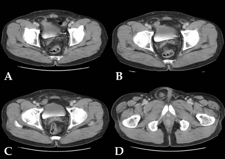
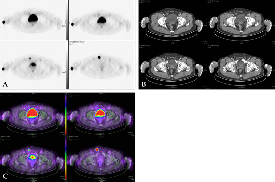
- A 70-year-old man with past history of hemicolectomy due to colon cancer underwent a follow-up abdominal/pelvic CT scan. CT revealed a right adrenal metastasis and then he underwent FDG-PET/CT study to search for other possible tumor recurrence. In PET images, other than right adrenal gland, there was an unexpected intense FDG uptake at right inguinal region and at first, it was considered to be an inguinal metastasis. However, correlation of PET images to concurrent CT data revealed it to be a bladder herniation. This case provides an example that analysis of PET images without corresponding CT images can lead to an insufficient interpretation or false positive diagnosis. Hence, radiologists should be aware of the importance of a combined analysis of PET and CT data in the interpretation of integrated PET/CT and rare but intriguing conditions, such as bladder herniation, during the evaluation of PET scans in colon cancer patients.
Keyword
MeSH Terms
-
Abdominal Neoplasms/radionuclide imaging/secondary
Aged
Colonic Neoplasms/pathology/radiography/radionuclide imaging
Diagnosis, Differential
False Positive Reactions
Fluorodeoxyglucose F18/*diagnostic use
Hernia, Inguinal/radiography/*radionuclide imaging
Humans
Male
*Positron-Emission Tomography
Radiopharmaceuticals/*diagnostic use
Tomography, X-Ray Computed
Urinary Bladder Diseases/radiography/*radionuclide imaging
Figure
Reference
-
1. Hustinx R. PET imaging in assessing gastrointestinal tumors. Radiol Clin North Am. 2004. 42:1123–1139. ix
Article2. Delbeke D, Martin WH. PET and PET-CT for evaluation of colorectal carcinoma. Semin Nucl Med. 2004. 34:209–223.
Article3. Huebner RH, Park KC, Shepherd JE, Schwimmer J, Czernin J, Phelps ME, et al. A meta-analysis of the literature for whole-body FDG PET detection of recurrent colorectal cancer. J Nucl Med. 2000. 41:1177–1189.4. Even-Sapir E, Parag Y, Lerman H, Gutman M, Levine C, Rabau M, et al. Detection of recurrence in patients with rectal cancer: PET/CT after abdominoperineal or anterior resection. Radiology. 2004. 232:815–822.
Article5. Conde Sánchez JM, Espinosa Olmedo J, Salazar Murillo R, Vega Toro P, Amaya Gutiérrez J, Alonso Flores J, et al. Giant inguino-scrotal hernia of the bladder. Clinical case and review of the literature. Actas Urol Esp. 2001. 25:315–319.6. Koontz AR. Sliding hernia of diverticulum of bladder. AMA Arch Surg. 1955. 70:436–438.
Article7. Fisher PC, Hollenbeck BK, Montgomery JS, Underwood W 3rd. Inguinal bladder hernia masking bowel ischemia. Urology. 2004. 63:175–176.
Article8. Ciancio G, Burke GW, Nery J, Huson H, Coker D, Miller J. Positional obstructive uropathy secondary to ureteroneocystostomy herniation in a renal transplant recipient. J Urol. 1995. 154:1471–1472.
Article9. Catalano O. Computed tomography findings in scrotal cystocele. Eur J Radiol. 1995. 21:126–127.
Article10. Andac N, Baltacioğlu F, Tüsney D, Cimşit NC, Ekinci G, Biren T. Inguinoscrotal bladder herniation: is CT a useful tool in diagnosis? Clin Imaging. 2002. 26:347–348.11. Pirson AS, Krug B, Lacrosse M, Luyx D, Barbeaux A, Borght TV. Bladder hernia simulating metastatic lesion on FDG PET study. Clin Nucl Med. 2004. 29:767.
Article12. Cohade C, Osman M, Leal J, Wahl RL. Direct comparison of (18)F-FDG PET and PET/CT in patients with colorectal carcinoma. J Nucl Med. 2003. 44:1797–1803.13. Gutman F, Alberini JL, Wartski M, Vilain D, Le Stanc E, Sarandi F, et al. Incidental colonic focal lesions detected by FDG PET/CT. AJR Am J Roentgenol. 2005. 185:495–500.
Article
- Full Text Links
- Actions
-
Cited
- CITED
-
- Close
- Share
- Similar articles
-
- Inguinal Herniation of Urinary Bladder on F-18 Sodium Fluoride (NaF) PET-CT
- Discordant Molecular Imaging Findings with 2‑[18F]FDG and [68Ga] Ga‑PSMA PET/CT in a Patient with Both Bladder and Prostate Cancer
- Usefulness of 18F-FDG PET/CT to Detect Metastatic Mucinous Adenocarcinoma Within an Inguinal Hernia
- Value of Bone Scan in Addition to F-18 FDG PET/CT and Characteristics of Discordant Lesions between F-18 FDG PET/CT and Bone Scan in the Spinal Bony Metastasis
- Massive Inguinal Bladder Hernia into the Scrotum


