Clinical Value of CT-Guided Needle Biopsy for Retroperitoneal Lesions
- Affiliations
-
- 1Department of Diagnostic and Interventional Radiology, Aichi Cancer Center Hospital, Nagoya 464-8681, Japan. 105824@aichi-cc.jp
- KMID: 1122332
- DOI: http://doi.org/10.3348/kjr.2011.12.3.351
Abstract
OBJECTIVE
The purpose of this study was to investigate retrospectively the clinical procedural performance of CT-guided needle biopsy for retroperitoneal lesions.
MATERIALS AND METHODS
CT-guided needle biopsy was performed in 74 consecutive patients (M:F = 44:30; mean age, 59.7 years) with retroperitoneal lesions between April 1998 and June 2009. The target lesion ranged from 1.5 to 12.5 cm in size. The biopsy access path ranged from 3.5 to 11.5 cm in depth. A biopsy specimen was obtained using an 18-gauge core needle under a CT or CT-fluoroscopy guidance and with the patient under local anesthesia. The histopathological diagnoses from the biopsies were obtained. The diagnostic confirmation of the subtype of lymphoma was evaluated.
RESULTS
Satisfactory biopsy samples were obtained in 73 (99%) of 74 patients and a pathological diagnosis was made in 70 (95%) of 74 patients. Sixty three lesions were malignant (45 lymphomas, nine primary tumors, nine lymph node metastases) and seven were benign. The subtype of lymphoma was specified in 43 (96%) of 45 patients who were diagnosed with lymphoma. Analysis of the value of CT-guided biopsy in this series indicated 63 true positives, zero false positive, six true negatives and five false negatives. This test had a sensitivity of 93%, a specificity of 100% and an accuracy of 93%. No major complications were seen and minor complications were noted in seven patients (five with local hematomas, two with transient pain at the puncture site).
CONCLUSION
CT-guided needle biopsy for retroperitoneal lesions is highly practical and useful, and particularly for determining the subtypes in patients with lymphoma.
MeSH Terms
Figure
-
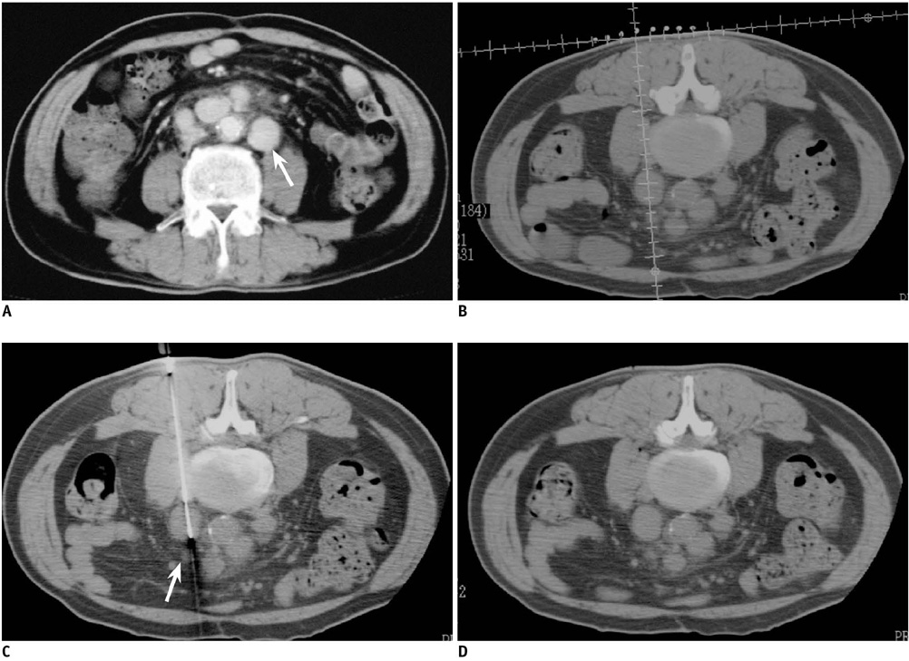
Fig. 1 53-year-old man with paraaortic lesion. A. Diagnostic axial contrast enhanced CT scan that was obtained prior to biopsy procedure with patient in supine position shows 2-cm paraaortic mass lesion (arrow). B. Initial axial CT scan obtained with patient in prone position shows tracking scale crossing retrocaval mass lesion. Distance from skin to leading edge of lesion was 10 cm. C. CT fluoroscopic image obtained during biopsy procedure shows safe insertion of biopsy needle (arrow) into lesion. Pathologic diagnosis was nodular sclerosis of Hodgkin's disease. D. CT scan immediately after two punctures reveals no complications.
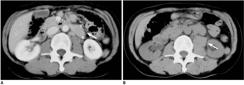
Fig. 2 56-year-old man with paraaortic lesion. A. Diagnostic axial contrast enhanced CT scan obtained prior to biopsy procedure with patient in supine position shows 3-cm paraaortic mass lesion (arrow). B. CT scan immediately after two punctures by posterior approach for biopsy reveals retroperitoneal hematoma (arrow).
Reference
-
1. Hopper KD. Percutaneous, radiographically guided biopsy: a history. Radiology. 1995. 196:329–333.2. Gazelle GS, Haaga JR. Guided percutaneous biopsy of intraabdominal lesions. AJR Am J Roentgenol. 1989. 153:929–935.3. Ben-Yehuda D, Polliack A, Okon E, Sherman Y, Fields S, Lebenshart P, et al. Image-guided core-needle biopsy in malignant lymphoma: experience with 100 patients that suggests the technique is reliable. J Clin Oncol. 1996. 14:2431–2434.4. Guimaraes AC, Chapchap P, de Camargo B, Chojniak R. Computed tomography-guided needle biopsies in pediatric oncology. J Pediatr Surg. 2003. 38:1066–1068.5. Husband JE, Golding SJ. The role of computed tomography-guided needle biopsy in an oncology service. Clin Radiol. 1983. 34:255–260.6. Sklair-Levy M, Polliack A, Shaham D, Applbaum YH, Gillis S, Ben-Yehuda D, et al. CT-guided core-needle biopsy in the diagnosis of mediastinal lymphoma. Eur Radiol. 2000. 10:714–718.7. Demharter J, Muller P, Wagner T, Schlimok G, Haude K, Bohndorf K. Percutaneous core-needle biopsy of enlarged lymph nodes in the diagnosis and subclassification of malignant lymphomas. Eur Radiol. 2001. 11:276–283.8. Pappa VI, Hussain HK, Reznek RH, Whelan J, Norton AJ, Wilson AM, et al. Role of image-guided core-needle biopsy in the management of patients with lymphoma. J Clin Oncol. 1996. 14:2427–2430.9. Irie T, Kajitani M, Matsueda K, Arai Y, Inaba Y, Kujiraoka Y, et al. Biopsy of lung nodules with use of I-I device under intermittent CT fluoroscopic guidance: preliminary clinical study. J Vasc Interv Radiol. 2001. 12:215–219.10. Welch TJ, Sheedy PF 2nd, Johnson CD, Johnson CM, Stephens DH. CT-guided biopsy: prospective analysis of 1,000 procedures. Radiology. 1989. 171:493–496.11. Chojniak R, Isberner RK, Viana LM, Yu LS, Aita AA, Soares FA. Computed tomography guided needle biopsy: experience from 1,300 procedures. Sao Paulo Med J. 2006. 124:10–14.12. Libicher M, Noldge G, Radeleff B, Gholipur F, Richter GM. Value of CT-guided biopsy in malignant lymphoma. Radiologe. 2002. 42:1009–1012.13. Knelson M, Haaga J, Lazarus H, Ghosh C, Abdul-Karim F, Sorenson K. Computed tomography-guided retroperitoneal biopsies. J Clin Oncol. 1989. 7:1169–1173.14. Agid R, Sklair-Levy M, Bloom AI, Lieberman S, Polliack A, Ben-Yehuda D, et al. CT-guided biopsy with cutting-edge needle for the diagnosis of malignant lymphoma: experience of 267 biopsies. Clin Radiol. 2003. 58:143–147.15. Stattaus J, Kalkmann J, Kuehl H, Metz KA, Nowrousian MR, Forsting M, et al. Diagnostic yield of computed tomography-guided coaxial core biopsy of undetermined masses in the free retroperitoneal space: single-center experience. Cardiovasc Intervent Radiol. 2008. 31:919–925.16. Carlson SK, Felmlee JP, Bender CE, Ehman RL, Classic KL, Hoskin TL, et al. CT fluoroscopy-guided biopsy of the lung or upper abdomen with a breath-hold monitoring and feedback system: a prospective randomized controlled clinical trial. Radiology. 2005. 237:701–708.17. Cheung JY, Kim Y, Shim SS, Lim SM. Combined fluoroscopy- and CT-guided transthoracic needle biopsy using a C-arm cone-beam CT system: comparison with fluoroscopy-guided biopsy. Korean J Radiol. 2011. 12:89–96.18. de Bazelaire C, Farges C, Mathieu O, Zagdanski AM, Bourrier P, Frija J, et al. Blunt-tip coaxial introducer: a revisited tool for difficult CT-guided biopsy in the chest and abdomen. AJR Am J Roentgenol. 2009. 193:W144–W148.19. Memel DS, Dodd GD 3rd, Esola CC. Efficacy of sonography as a guidance technique for biopsy of abdominal, pelvic, and retroperitoneal lymph nodes. AJR Am J Roentgenol. 1996. 167:957–962.20. al-Mofleh IA. Ultrasound-guided fine needle aspiration of retroperitoneal, abdominal and pelvic lymph nodes. Diagnostic reliability. Acta Cytol. 1992. 36:413–441.21. Adam G, Bucker A, Nolte-Ernsting C, Tacke J, Gunther RW. Interventional MR imaging: percutaneous abdominal and skeletal biopsies and drainages of the abdomen. Eur Radiol. 1999. 9:1471–1478.22. Lee MH, Lufkin RB, Borges A, Lu DS, Sinha S, Farahani K, et al. MR-guided procedures using contemporaneous imaging frameless stereotaxis in an open-configuration system. J Comput Assist Tomogr. 1998. 22:998–1005.23. Kariniemi J, Blanco Sequeiros R, Ojala R, Tervonen O. MRI-guided abdominal biopsy in a 0.23-T open-configuration MRI system. Eur Radiol. 2005. 15:1256–1126.24. Zangos S, Eichler K, Wetter A, Lehnert T, Hammerstingl R, Diebold T, et al. MR-guided biopsies of lesions in the retroperitoneal space: technique and results. Eur Radiol. 2006. 16:307–312.25. Alanen J, Keski-Nisula L, Blanco-Sequeiros R, Tervonen O. Cost comparison analysis of low-field (0.23 T) MRI- and CT-guided bone biopsies. Eur Radiol. 2004. 14:123–112.
- Full Text Links
-
- Actions
-
Cited
- CITED
-
- Close
- Share
-
- Similar articles
-
- CT-Guided Biopsy of Pulmonary Lesions: A Comparison of Diagnostic Accuracy and Complication Rate betweenAutomated Gun Biopsy and Fine Needle Aspiration Biopsy
- CT-guided bone biopsy
- CT-guided fine needle biopsy of intraorbital masses
- CT-guided percutaneous fine-needle aspiration biopsy of thoracic and retroperitoneal lesion
- A Simplified Method by CT Guided Needle Placement of Intracranial Lesions Using U-Loop
