Hepatic Metastasis from Choriocarcinoma: Angiographic Findings in Two Cases
- Affiliations
-
- 1Department of Diagnostic Radiology, Kyung Hee University College of Medicine, Korea. ohjh6108@hanmail.net
- 2Department of Internal Medicine, Kyung Hee University College of Medicine, Korea.
- KMID: 1118794
- DOI: http://doi.org/10.3348/kjr.2002.3.4.260
Abstract
- We report two cases of hepatic metastases from choriocarcinoma in women of childbearing age in whom imaging studies performed at presentation revealed the presence of liver masses, and who had clinically progressive anemia or intraabdominal hemorrhage. CT demonstrated heterogeneously enhanced liver masses. Characteristic angiographic findings included hypervascular hepatic masses with aneurysmal dilatations of the peripheral hepatic arteries at the arterial phase and persistent vascular lakes at the venous phase.
MeSH Terms
Figure
-
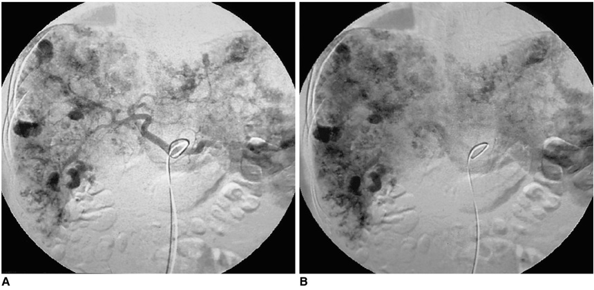
Fig. 1 Case 1. Hepatic metastasis from choriocarcinoma in a 33-year-old woman. A. Arterial-phase celiac angiogram depicts multiple hypervascular masses with abnormal tumor vessels and saccular aneurysmal dilatation of the peripheral end of hepatic arteries in the liver and spleen. B. Venous phase celiac angiogram demonstrates aneurysmal hepatic and splenic vascular lakes, but active bleeding is not apparent.
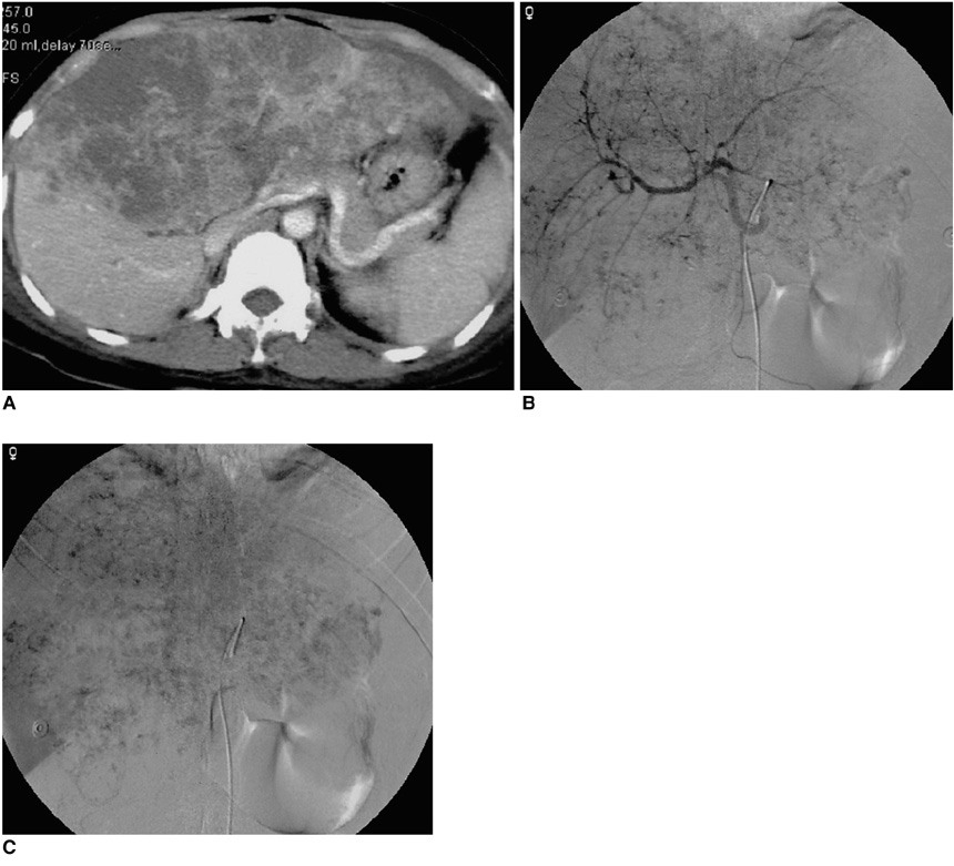
Fig. 2 Case 2. Hepatic metastasis from choriocarcinoma in a 33-year-old woman. A. Contrast-enhanced CT image depicts huge, ill-defined, heterogeneously enhanced masses in both lobes of the liver, and thrombosis in the left portal vein. B. Arterial-phase hepatic arteriogram reveals huge, multiple, exophytic hypervascular masses with saccular aneurysmal dilatation of the peripheral hepatic arteries and arteriovenous shunting in both lobes of the liver. C. Venous-phase hepatic arteriogram demonstrates persistent hepatic vascular lakes.
Reference
-
1. Hillard AE, Allen RW, Beale G. Metastatic choriocarcinoma: correlation of MRI, CT, and angiography. South Med J. 1993. 86:1299–1302.2. Green CL, Angtuaco TL, Shah HR, Parmley TH. Gestational trophoblastic disease: a spectrum of radiologic diagnosis. RadioGraphics. 1996. 16:1371–1384.3. Alveyn CG, Loehry CA. Hepatic metastases due to choriocarcinoma. Postgrad Med J. 1988. 64:941–942.4. Heatom GE, Matthews TH, Christopherson WM. Malignant trophoblastic tumors with massive hemorrhage presenting as liver primary: a report of two cases. Am J Surg Pathol. 1986. 10:342–347.5. Erb RE, Gibler WB. Massive hemoperitoneum following rupture of hepatic metastases from unsuspected choriocarcinoma. Am J Emerg Med. 1989. 7:196–198.6. Watanabe AS, Smoker WR. Computed tomography and angiographic findings in metastatic choriocarcinoma. J Comput Assist Tomogr. 1989. 13:319–322.7. Komeichi T, Igarashi K, Takigami M, et al. A case of metastatic choriocarcinoma associated with cerebral thrombosis and aneurysmal formation. No Shinkei Geka. 1996. 24:463–467.8. Yang DM, Yoon MH, Kim HS, Kim HS, Shin DB. Intrarenal pseudoaneurysms complicating renal choriocarcinoma metastases: treatment with coil embolization. Clin Imaging. 2000. 24:217–220.
- Full Text Links
-
- Actions
-
Cited
- CITED
-
- Close
- Share
-
- Similar articles
-
- Choriocarcinoma in Kidney : A Report of Two Cases
- Angiographic findings of choriocarcinoma
- Primary Hepatic Choriocarcinoma: A Case Report
- Two Cases of Surgical Intervention in Persistent Localized Choriocarcinoma
- Composite Adenocarcinoma and Choriocarcinoma of the Sigmoid Colon with Hepatic Metastasis of the Choriocarcinomatous Component
