Korean J Radiol.
2002 Dec;3(4):219-228. 10.3348/kjr.2002.3.4.219.
Intracranial and Spinal Ependymomas: Review of MR Images in 61 Patients
- Affiliations
-
- 1Department of Radiology, Seoul National University College of Medicine; Institute of Radiation Medicine, SNUMRC; Clinical Research Institute, Seoul National University Hospital, Korea. changkh@snu.ac.kr
- KMID: 1118788
- DOI: http://doi.org/10.3348/kjr.2002.3.4.219
Abstract
OBJECTIVE
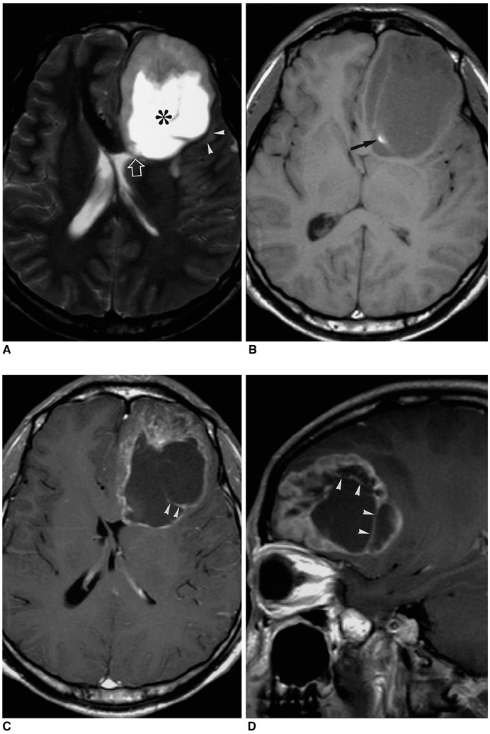
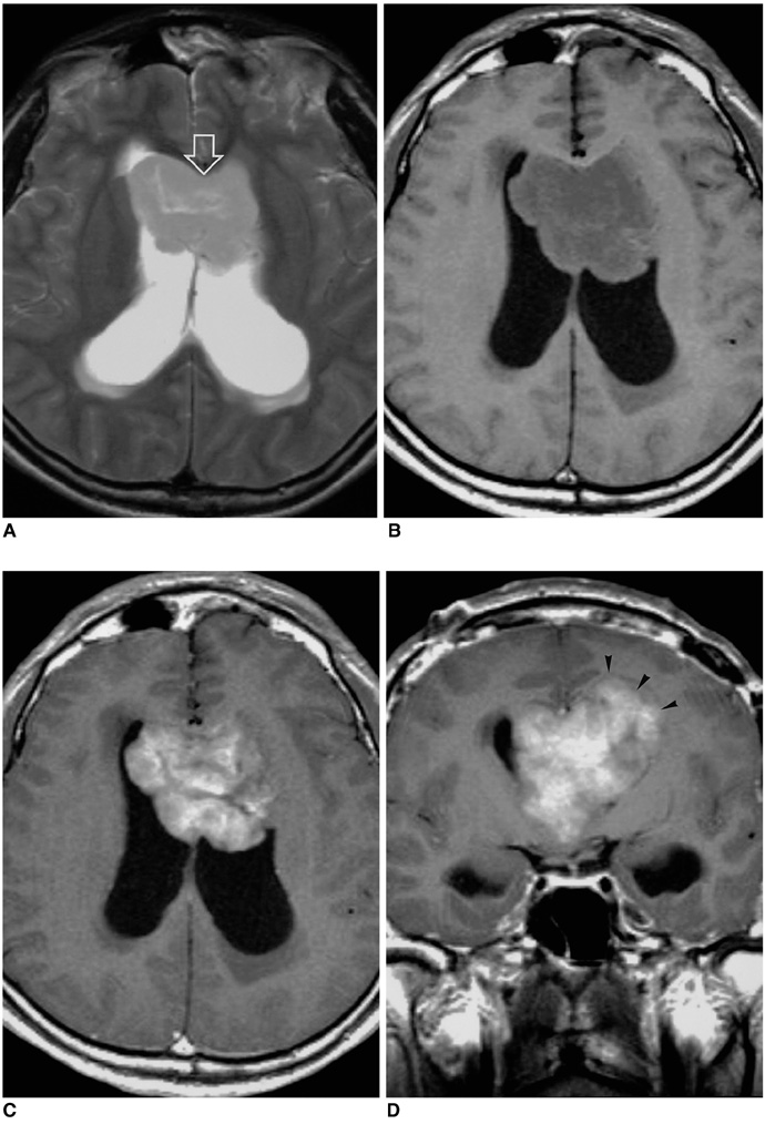
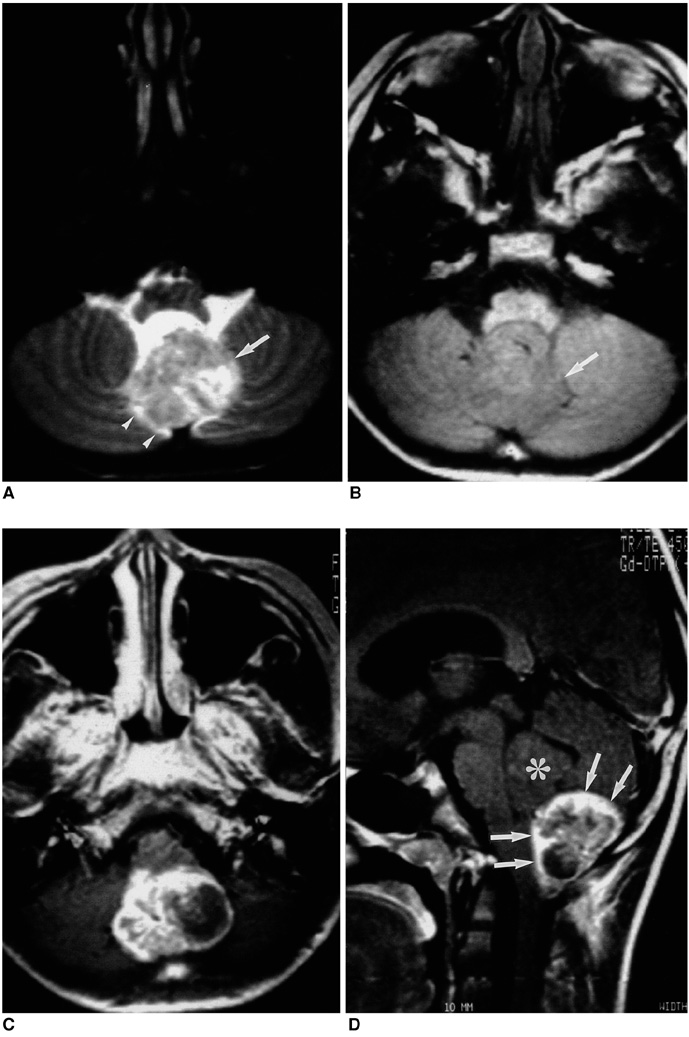
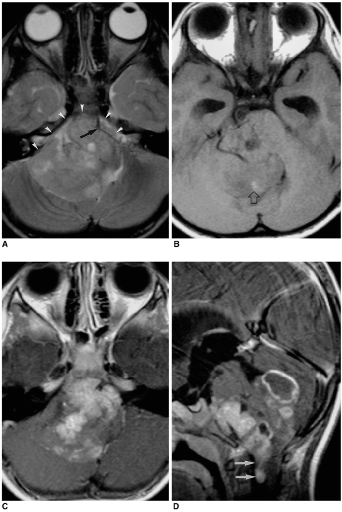
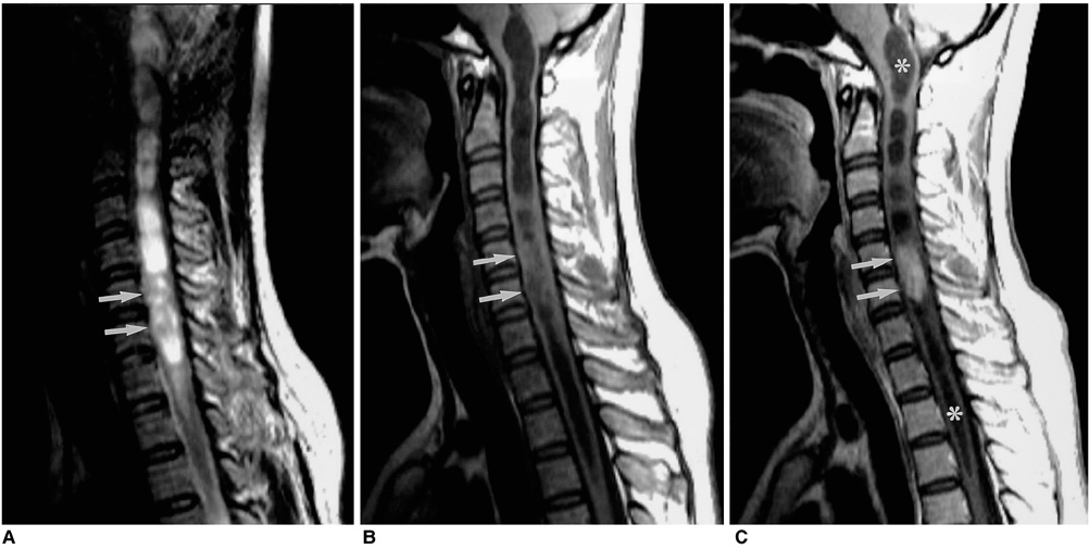
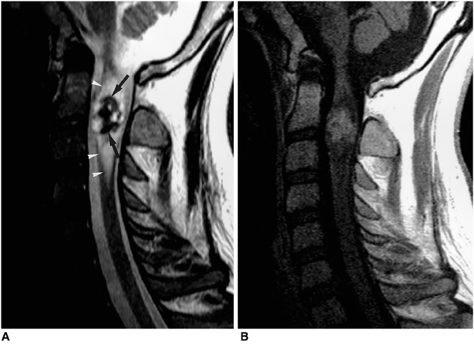
To compare the age distribution and characteristic MR imaging findings of ependymoma for each typical location within the neuraxis. MATERIALS AND METHODS: During a recent eleven-year period, MR images of 61 patients with histologically proven ependymomas were obtained and retrospectively reviewed in terms of incidence, peak age, location, size, signal intensity, the presence or absence of cyst and hemorrhage, enhancement pattern, and other associated findings. RESULTS: Among the 61 patients, tumor location was spinal in 35 (57%), infrartentorial in 19 (31%), and supratentorial in seven (12%). In four of these seven, the tumor was located in brain parenchyma, and in most cases developed between the third and fifth decade. Approximately half of the infratentorial tumors occurred during the first decade. The signal intensity of ependymomas was nonspecific, regardless of their location. A cystic component was seen in 71% (5/7) of supratentorial, 74% (14/19) of infratentorial, and 14% (5/35) of spinal cord tumors. Forty- nine percent (17/35) of those in the spinal cord were associated with rostral and/or caudal reactive cysts. Intratumoral hemorrhage occurred in 57% (4/7) of supratentorial, 32% (6/19) of infratentorial, and 9% (3/35) of spinal cord tumors. In 17% (6/35) of spinal ependymomas, a curvilinear low T2 signal, suggesting marginal hemorrhage, was seen at the upper and/or lower margins of the tumors. Peritumoral edema occurred in 57% (4/7) of supratentorial, 16% (3/19) of infratentorial and 23% (8/35) of spinal cord tumors. Seventy-two percent (5/7) of supratentorial and 95% (18/19) of infratentorial tumors showed heterogeneous enhancement, while in 50% (17/34) of spinal cord tumors, enhancement was homogeneous. CONCLUSION: Even though the MR imaging findings of ependymomas vary and are nonspecific, awareness of these findings, and of tumor distribution according to age, is helpful and increases the likelihood of correct preoperative clinical diagnosis.
MeSH Terms
Figure
Reference
-
1. Russel DS, Rubenstein LJ. Pathology of tumors of the nervous system. 1989. 5th ed. Baltimore: Williams & Wilkins;192–219.2. Rezai AR, Woo HH, Lee M, Cohen H, Zagzag D, Epstein FJ. Disseminated ependymomas of the central nervous system. J Neurosurg. 1996. 85:618–624.3. Fokes EC, Earle KM. Ependymomas: clinical and pathological aspects. J Neurosurg. 1969. 30:585–594.4. Lefton DR, Pinto RS, Martin SW. MRI features of intracranial and spinal ependymomas. Pediatr Neurosurg. 1998. 28:97–105.5. Fine MJ, Kricheff II, Freed D, Epstein FJ. Spinal cord ependymomas: MR imaging features. Radiology. 1995. 197:655–658.6. Kahan H, Sklar EM, Post MJ, Bruce JH. MR characteristics of histopathologic subtypes of spinal ependymoma. AJNR Am J Neuroradiol. 1996. 17:143–150.7. Greenfield JG. Greenfield's Neuropathology. 1992. 6th ed. New York: Oxford University Press;636–644.8. Barone BM, Elvidge AR. Ependymoma: a clinical survey. J Neurosurg. 1970. 33:428–438.9. Kernhan JW, Sayre GP. Tumors of the central nervous system: Atlas of tumor pathology Section X, Fascicle 35. 1952. Washington: Armed Forces Institute of Pathology.10. Armington WG, Osborn AG, Cubberley DA, et al. Supratentorial ependymoma: CT appearance. Radiology. 1985. 157:367–372.11. Swartz JD, Zimmerman RA, Bilaniuk LT. Computed tomography of intracranial ependymomas. Radiology. 1982. 143:97–101.12. Svien HJ, Mabon RF, Kernohan JW, Craig W. Ependymoma of the brain: pathologic aspects. Neurology. 1953. 3:1–15.13. Tortori-Donati P, Fondelli MP, Cama A, Garre ML, Rossi A, Andreussi L. Ependymomas of the posterior cranial fossa: CT and MRI findings. Neuroradiology. 1995. 37(3):238–243.14. Lee BCP, Kneeland JB, Cahill PT, Deck MDF. MR recognition of supratentorial tumors. AJNR Am J Neuroradiol. 1985. 6:871–878.15. Komiyama M, Yagura H, Baba M, et al. MR imaging: possibility of tissue characterization of brain tumors using T1 and T2 values. AJNR Am J Neuroradiol. 1987. 8:65–70.16. Coulon RA, Till K. Intracranial ependymomas in children: a review of 43 cases. Child's Brain. 1977. 3:154–168.17. Furie DM, Provenzale JM. Supratentorial ependymomas and subependymomas: CT and MR appearance. J Comput Assist Tomogr. 1995. 19:518–526.18. Li MH, Holtas S. MR imaging of spinal intramedullary tumors. Acta Radiol. 1991. 32:505–513.19. Goy AMC, Pinto RS, Raghavendra BN, Epstein FJ, Kricheff II. Intramedullary spinal cord tumors: MR imaging, with emphasis on associated cysts. Radiology. 1986. 161:381–386.20. Epstein FJ, Farmer JP, Freed D. Adult intramedullary spinal cord ependymomas: the result of surgery in 38 patients. J Neurosurg. 1993. 79:204–209.21. Naidich TP, Zimmerman RA. Primary brain tumors in children. Semin Roentgenol. 1984. 19:100–114.22. Nemoto Y, Inoue Y, Tashiro T, et al. Intramedullary spinal cord tumors: significance of associated hemorrhage at MR imaging. Radiology. 1992. 182:793–796.23. Parizel PM, Bakeriaux D, Rodesch G, et al. Gd-DTPA-enhanced MR imaging of spinal tumors. AJR Am J Roentgenol. 1989. 152:1087–1096.24. Johnson PC, Hunt SJ, Drayer BP. Human cerebral gliomas: correlation of postmortem MR imaging and neuropathologic findings. Radiology. 1989. 170:211–217.
- Full Text Links
- Actions
-
Cited
- CITED
-
- Close
- Share
- Similar articles
-
- MR Imaging of Intramedullary Tumors of the Spinal Cord
- Clinicoradiologic Characteristics of Intradural Extramedullary Conventional Spinal Ependymoma
- A Case of Giant Ependymomas of the Cauda Equina associated with Thoracic Intradural Spinal A-V Malformations
- Intradural Extramedullary Ependymoma with Hemorrhage: A Case Report
- The Similarities and Differences between Intracranial and Spinal Ependymomas : A Review from a Genetic Research Perspective

