The Radiological Spectrum of Orbital Pathologies that Involve the Lacrimal Gland and the Lacrimal Fossa
- Affiliations
-
- 1Department of Radiology, St Mary's Hospital, Catholic University College of Medicine, Seoul, Korea. ahn-kj@catholic.ac.kr
- 2Department of Ophthalmology, St Mary's Hospital, Catholic University College of Medicine, Seoul, Korea.
- KMID: 1110731
- DOI: http://doi.org/10.3348/kjr.2007.8.4.336
Abstract
- CT and MRI are utilized to differentiate between different types of masses and to determine the extent of lesions involving the lacrimal gland and the fossa. Although many diseases that affect the lacrimal gland and fossa are specifically diagnosed by imaging, it is frequently very difficult to differentiate each specific disease on the basis of image characteristics alone due to intrinsic similarities. In lacrimal gland epithelial tumors, benign pleomorphic adenomas are seen most commonly with a well defined benign appearance, and a malignant adenoid cystic carcinoma is seen with a typical invasive malignant appearance. However, a malignant myoepithelial carcinoma is seen with a benign looking appearance. Lymphomatous lesions of the lacrimal gland include a broad spectrum ranging from reactive hyperplasia to malignant lymphoma. These lesions can be very difficult to differentiate both radiologically and pathologically. Generally, lymphomas tend to occur in older patients. The developmental cystic lesions found in the lacrimal fossa such as dermoid and epidermoid cysts can be diagnosed when the cyst involves the superior temporal quadrant of the orbit and manifests as a non-enhancing cystic mass and, in case of a lipoma, it is diagnosed as a total fatty mass. However, masses of granulocytic sarcoma and xanthogranuloma, as well as vascular masses, such as a hemangiopericytoma, are difficult to diagnose correctly on the basis of preoperative imaging findings alone. A careful clinical evaluation and moreover, a pathologic verification, are needed. In this pictorial review, the various imaging spectrums of pathologic masses involving the lacrimal gland and fossa are presented, along with appropriate anatomy and pathology reviews.
MeSH Terms
-
Carcinoma, Squamous Cell/radiography
Conjunctival Neoplasms/radiography
Cysts/radiography
Eye Neoplasms/*radiography
Hemangiopericytoma/radiography
Humans
Lacrimal Apparatus/*pathology
Lacrimal Apparatus Diseases/radiography
Lipoma/radiography
Lymphoma/radiography
Neoplasms, Glandular and Epithelial/radiography
Neurofibroma/radiography
Sarcoma, Myeloid/radiography
Figure
-
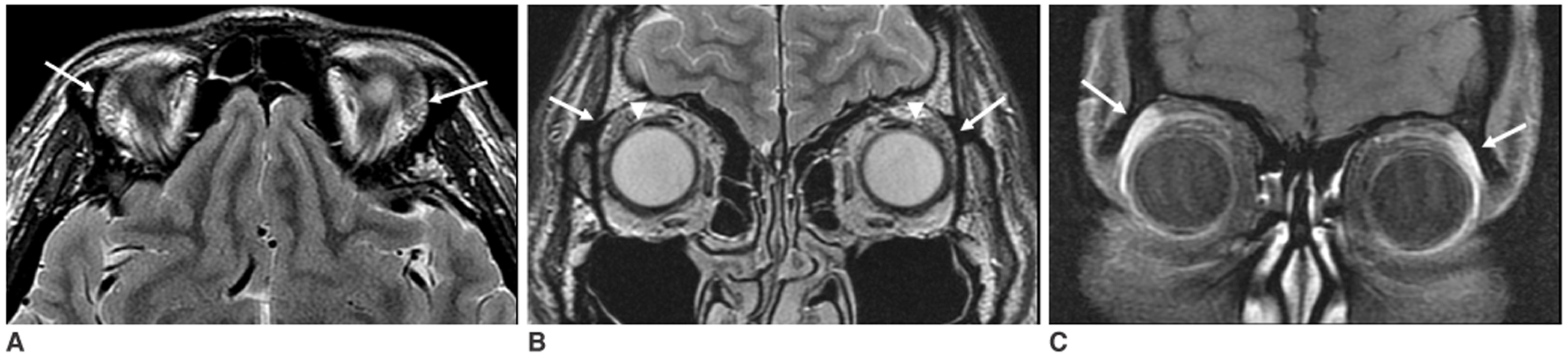
Fig. 1 The normal anatomy of the lacrimal gland and fossa. A. A transverse T2-weighted MR image at the level of the lacrimal gland shows almond-shaped lacrimal glands at the superior lateral aspect of both eyeglobes (arrows). B. A coronal T2-weighted MR image at the level of the lacrimal gland and aponeurosis of the levator palpebrae muscle shows continuous extension of the aponeurosis into the lacrimal gland, which divides the gland into anterior palpebral and deeper orbital lobes (arrowheads). C. A coronal enhanced T1-weighted MR image shows well-enhanced lacrimal glands (arrows).
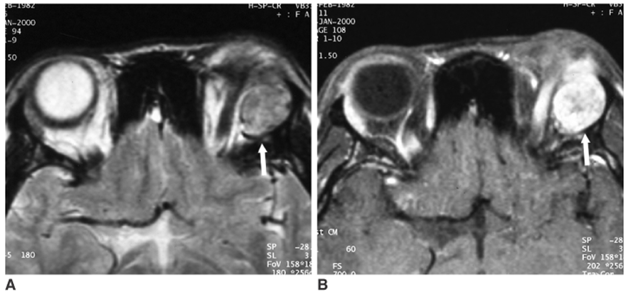
Fig. 2 A pleomorphic adenoma in a 44-year-old man. A. A well-circumscribed, spherical tumor is seen in the left lacrimal fossa with heterogeneous intermediate signal intensity on a T2-weighted image (arrow). B. On a gadolinium enhanced T1-weighted image, strong enhancement is noted in the mass (arrow).
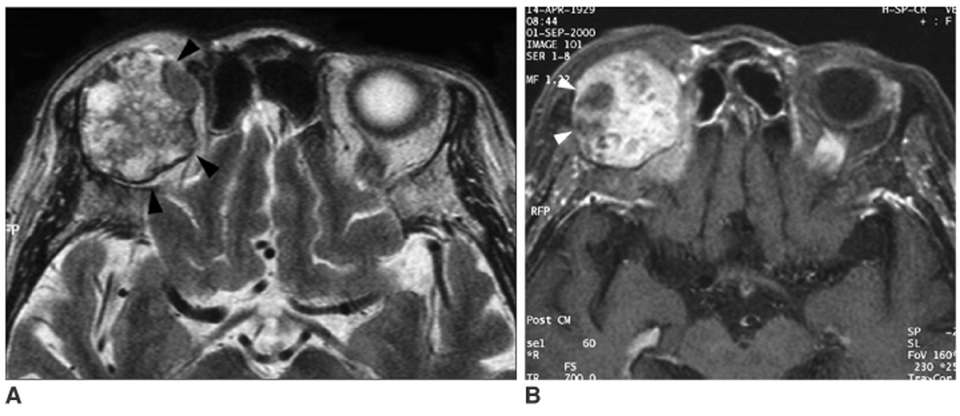
Fig. 3 A pleomorphic adenoma in a 63-year-old woman. A. A well-circumscribed mass is seen in the right lacrimal fossa with heterogenous high signal intensity on T2-weighted images (arrowheads). B. On a gadolinium enhanced T1-weighted image, the tumor shows strong enhancement with poorly enhanced areas (arrowheads), representing cystic changes.
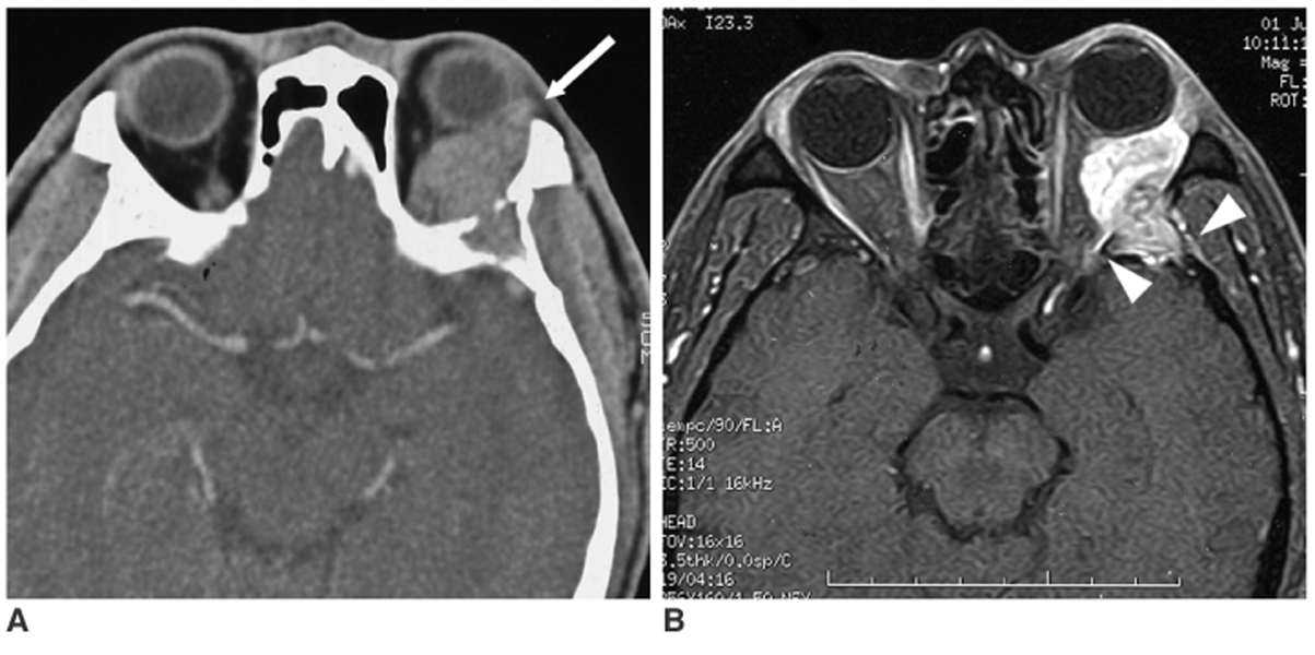
Fig. 4 An adenoid cystic carcinoma in a 55-year-old man. A. Contrast enhanced CT scan shows a well-enhancing soft tissue mass (arrow) in the left lacrimal fossa with destruction of the lateral orbital wall. B. A gadolinium enhanced T1-weighted image shows intracranial extension into the middle cranial fossa with dural enhancement (arrowheads).
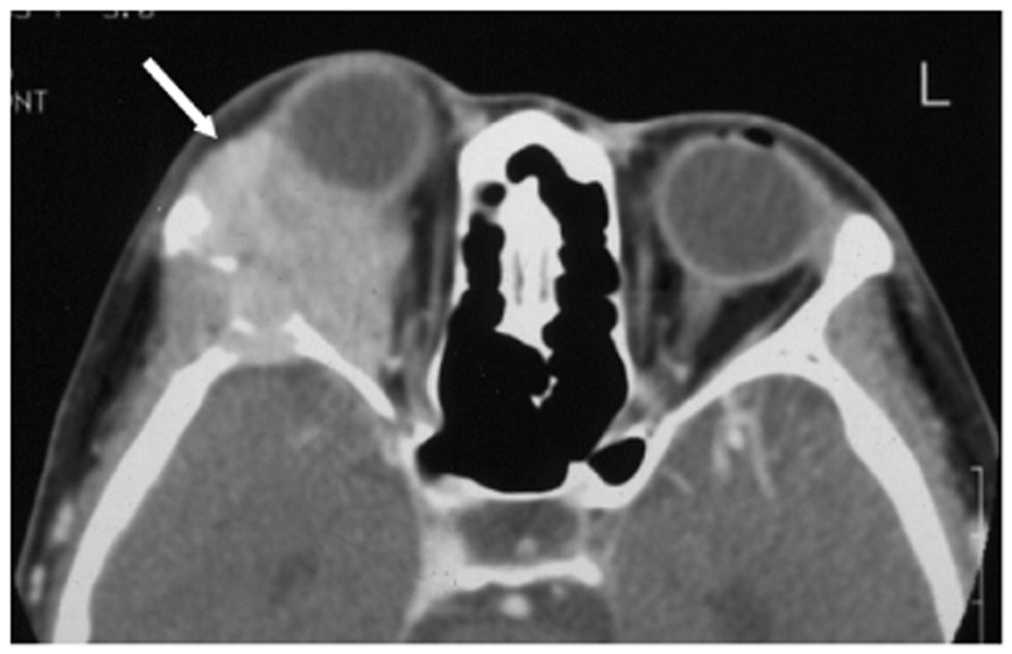
Fig. 5 A myoepithelial carcinoma in a 32-year-old man. A contrast enhanced CT scan shows a well-enhancing tumor (arrow) in the right lacrimal fossa with bony destruction of zygomatic and sphenoid bone.
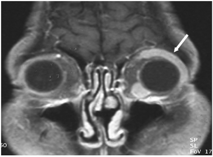
Fig. 6 A malignant lymphoma in a 41-year-old woman. A gadolinium enhanced T1-weighted coronal image shows an elongated soft tissue mass (arrow) along the superior lateral aspect of the left eye globe. This lesion shows homogeneous and strong enhancement. Another enhancing nodule is noted at the inferior medial aspect of the left eye globe.

Fig. 7 Lymphoid hyperplasia in a 43-year-old man. A contrast enhanced coronal CT scan shows a homogeneously-enhancing elongated mass involving the right lacrimal gland (arrow).
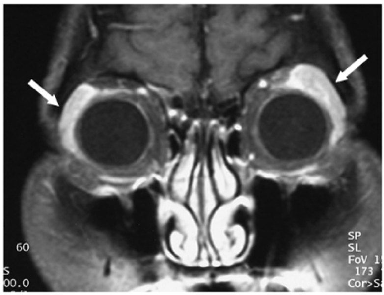
Fig. 8 Chronic dacryoadenitis in a 39-year-old woman. A gadolinium-enhanced T1-weighted coronal image shows diffuse and elongated enlargement of both lacrimal glands with contrast enhancement (arrows).

Fig. 9 An inflammatory pseudotumor in a 54-year-old woman. A contrast-enhanced CT scan shows an ill-defined enhancing mass in the superior lateral aspect of left eye globe (arrow). Subcutaneous infiltrations (arrowheads) are accompanied in the lateral outside of the left orbit.
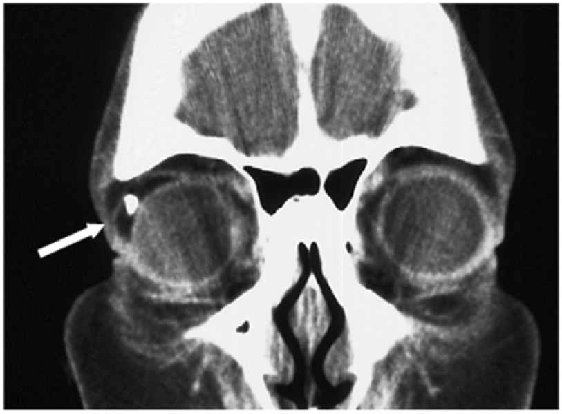
Fig. 10 A dermoid cyst in a 49-year-old woman. A coronal CT scan shows a fat-containing mass with nodular calcification in the superior lateral aspect of right orbit (arrow).
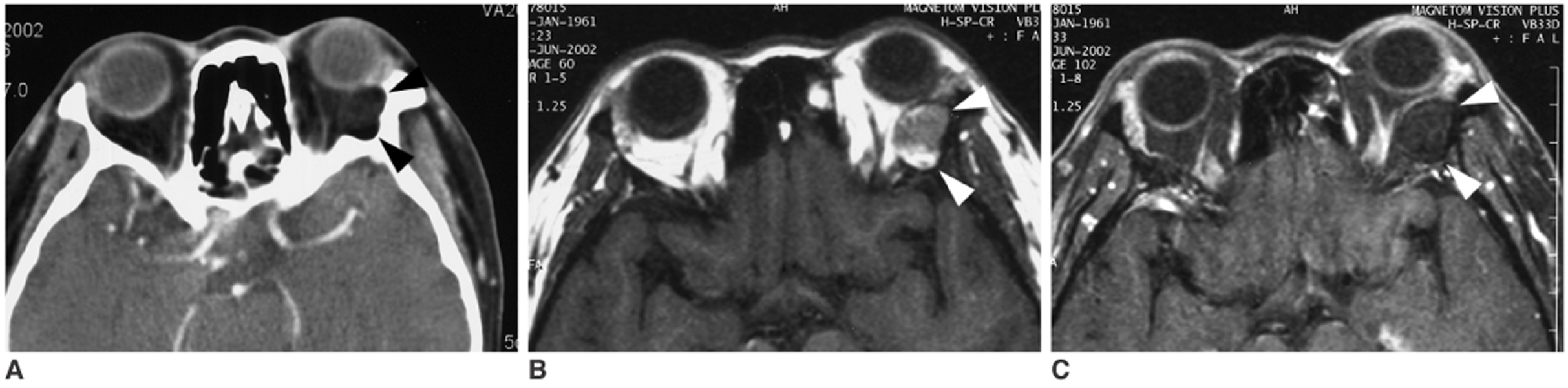
Fig. 11 An epidermoid cyst in a 35-year-old man. A. A contrast enhanced axial CT scan shows a fat-containing mass with bony scalloping at the lateral orbital wall (arrowheads). B. On a T1-weighted axial image, this mass shows heterogeneous intermediate to high signal intensity (arrowheads). C. On a contrast-enhanced, fat-suppressed T1-weighted image, no enhancement is noted in the mass (arrowheads).
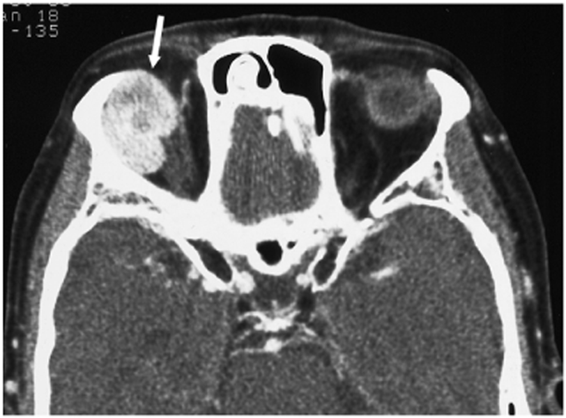
Fig. 12 A hemangiopericytoma in a 65-year-old man. A contrast-enhanced axial CT scan shows a strongly enhancing mass with a lobulated contour in the superior lateral aspect of the right orbit (arrow).
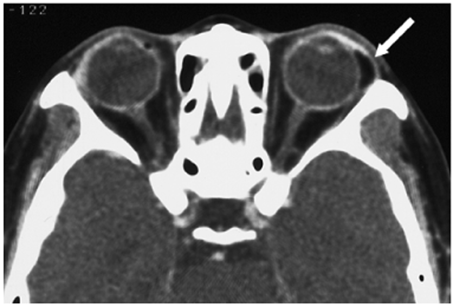
Fig. 13 A lipoma in a 64-year-old man. An axial CT scan shows a fat-containing mass at the superior lateral aspect of the left orbit (arrow).
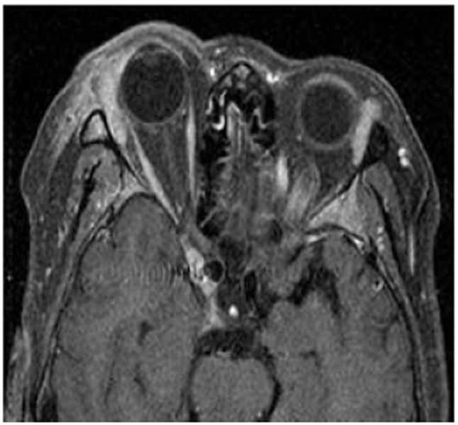
Fig. 14 A granulocytic sarcoma from chronic myeloid leukemia in a 40-year-old man. A gadolinium-enhanced T1-weighted axial image shows bilaterally-enlongated masses with strong enhancement in the superior lateral aspect of both orbits. Soft tissue infiltrations are noted around the right orbit mass.
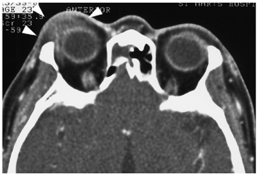
Fig. 15 Xanthogranuloma in a 62-year-old woman. A contrast enhanced axial CT scan shows poorly enhancing, soft tissue density infiltrations at the anterior lateral aspect of the right orbit (arrowheads).
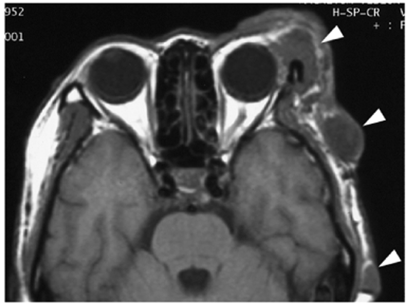
Fig. 16 Plexiform neurofibromatosis in a 66-year-old man. The T1-weighted axial image shows multiple lobulating masses involving the left lacrimal fossa and the subcutaneous layer of the left temporal region (arrowheads).
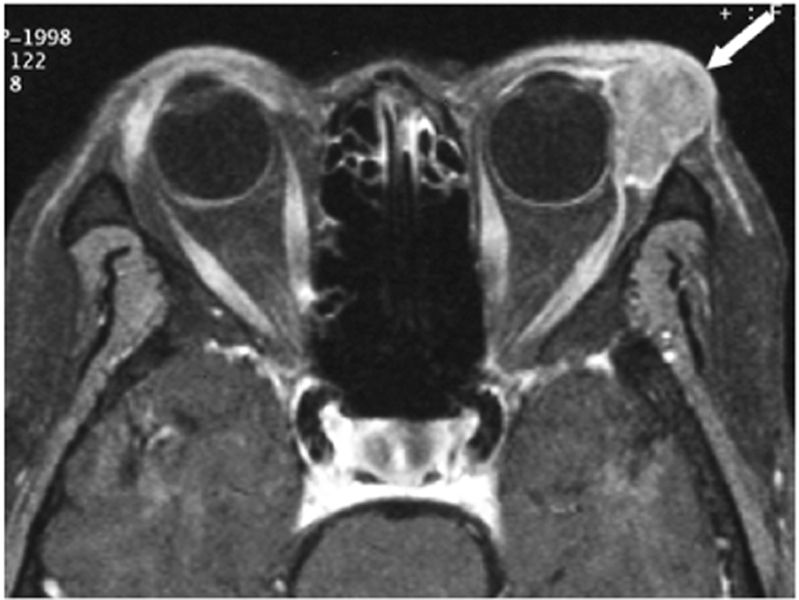
Fig. 17 Squamous cell carcinoma of the conjunctiva in an upper eyelid of a 61-year-old man. A gadolinium enhanced T1-weighted axial image shows a well-enhanced lobulated mass in the superior lateral aspect of the left eye globe (arrow). The thickened skin is not separable from the mass.
Cited by 1 articles
-
Calculated Brain CT Angiography Volumes of Lacrimal Glands in Normal Korean Orbits
Seoung Hyun An, Sang Wook Jin, Won Seok Yang, Hee Bae Ahn
J Korean Ophthalmol Soc. 2014;55(10):1413-1417. doi: 10.3341/jkos.2014.55.10.1413.
Reference
-
1. Shields CL, Shields JA, Eagle RC, Rathmell JP. Clinicopathologic review of 142 cases of lacrimal gland lesions. Ophthalmology. 1989. 96:431–435.2. Balchunas WR, Quencer RM, Byrne SF. Lacrimal Gland and Fossa Masses: evaluation by computed tomography and A-mode echography. Radiology. 1983. 149:751–758.3. Hesselink JR, Davis KR, Dallow RL, Roberson GH, Taveras JM. Computed tomography of masses in the lacrimal gland region. Radiology. 1979. 131:143–147.4. Mafee MF. Som PM, editor. Orbit: embryology, anatomy, and pathology. Head and neck imaging. 2003. 2nd ed. St. Louis: Mosby;529–654.5. Mizokami H, Inokuchi A, Sawatsubashi M, Takagi S, Tsuda K, Tokunaga O. Adenoid cystic carcinoma of the lacrimal gland with wide and severe myoepithelial differentiation. Auris Nasus Larynx. 2002. 29:77–82.6. Mafee MF, Haik BG. Lacrimal gland and fossa lesions: role of computed tomography. Radiol Clin North Am. 1987. 25:767–779.7. Narla LD, Newman B, Spottswood SS, Narla S, Kolli R. Inflammatory pseudotumor. Radiographics. 2003. 23:719–729.8. Valvassori GE, Sabnis SS, Mafee RF, Brown MS, Putterman A. Imaging of orbital lymphoproliferative disorders. Radiol Clin North Am. 1999. 37:135–150.9. Miszkiel KA, Sohaib SA, Rose GE, Cree IA, Moseley IF. Radiological and clinicopathological features of orbital xanthogranuloma. Br J Ophthalmol. 2000. 84:251–258.
- Full Text Links
-
- Actions
-
Cited
- CITED
-
- Close
- Share
-
- Similar articles
-
- A Case of Intraorbital Ectopic Lacrimal Gland with Lacrimal Duct
- A Case of Mucoepidermoid Carcinoma of the Lacrimal Gland
- Study of the Lacrimal Gland Size in Korean Using Orbital Computed Tomography
- The Change of Thickness of the Lacrimal Gland by Orbital Computed Tomographies associated with Aging
- Apocrine Hidrocystoma Presenting as a Lacrimal Gland Mass
