Does Ultrasound-Guided Directional Vacuum-Assisted Removal Help Eliminate Abnormal Nipple Discharge in Patients with Benign Intraductal Single Mass?
- Affiliations
-
- 1Department of Radiology and Clinical Research Institute, Seoul National University Hospital and the Institute of Radiation Medicine, Seoul National University Medical Research Center, Seoul 110-744, Korea. nariya@radiol.snu.ac.kr
- 2Department of Radiology, Hanyang University College of Medicine, Hanyang University Hospital, Seoul 133-792, Korea.
- 3Department of Radiology, Seoul National University Boramae Hospital, Seoul 156-012, Korea.
- 4Department of Radiology, Seoul National University Bundang Hospital, Gyeonggi-do 463-707, Korea.
- KMID: 1102560
- DOI: http://doi.org/10.3348/kjr.2009.10.6.575
Abstract
OBJECTIVE
To evaluate whether the removal of an intraductal mass using an ultrasound (US)-guided directional vacuum-assisted device can eliminate symptoms in patients presenting with abnormal nipple discharge. MATERIALS AND METHODS: Between March 2004 and October 2006, 36 patients who presented with abnormal nipple discharge, underwent US-guided, 11-gauge vacuum-assisted biopsy for a benign intraductal single mass on US. The ability of the procedure to eliminate nipple discharge was evaluated by physical examination during follow-up US. Lesion characteristics, biopsy variables, and histologic features were analyzed to identify factors affecting symptom resolution. RESULTS: Of the 36 lesions, 25 (69%) were intraductal papillomas, 10 (28%) were fibrocystic changes, and one (3%) was a fibroadenoma. The nipple discharge disappeared in 69% (25 of 36) of the women at a mean follow-up time of 25 months (range 12-42 month). There was no difference in the lesion characteristics, biopsy variables, and the histologic features between groups that eliminated the symptom compared those with persistent nipple discharge. CONCLUSION: US-guided directional vacuum-assisted removal of an intraductal mass appears to eliminate nipple discharge in only 69% of patients and thus, it should not be considered as an alternative to surgical excision.
MeSH Terms
-
Adult
Biopsy/*methods
Breast Neoplasms/pathology/*ultrasonography
Chi-Square Distribution
Exudates and Transudates/*ultrasonography
Female
Humans
Mammography
Middle Aged
Nipples/pathology/*ultrasonography
Papilloma, Intraductal/pathology/*ultrasonography
Retrospective Studies
*Ultrasonography, Interventional
*Ultrasonography, Mammary
Vacuum
Figure
-
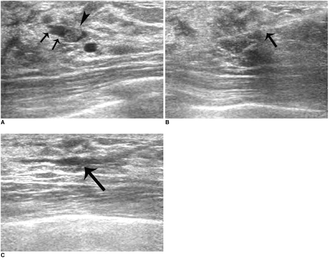
Fig. 1 44-year-old woman successfully treated for bloody nipple discharge by US-guided directional vacuum-assisted removal.A. Sonogram showing well-defined, intraductal single mass (arrowhead) within dilated duct (arrows).B. Sonogram showing directional vacuum-assisted probe (arrow) underneath lesion.C. Sonogram obtained immediately after US-guided directional vacuum-assisted removal revealing mild fluid collection within dilated duct (arrow). Mass was confirmed to be intraductal papilloma. No residual nipple discharge was apparent at six month follow-up.
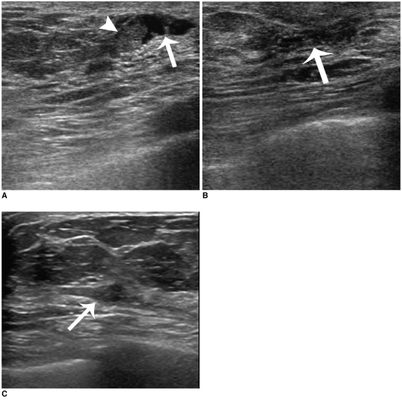
Fig. 2 48-year-old woman with remaining bloody nipple discharge symptom after US-guided directional vacuum-assisted removal.A. Sonogram showing well-defined, single intraductal mass (arrowhead) within dilated duct (arrow).B. Sonogram obtained immediately after US-guided directional vacuum-assisted removal revealing mild fluid collection within slightly dilated duct (arrow). Mass was confirmed as intraductal papilloma.C. Follow-up sonogram six months after US-guided directional vacuum-assisted removal showing low echoic mass (arrow) in subareolar area. Nipple discharge persisted and biopsy revealed recurrent intraductal papilloma.
Reference
-
1. Paterok EM, Rosenthal H, Säbel M. Nipple discharge and abnormal galactogram. Results of a long-term study (1964-1990). Eur J Obstet Gynecol Reprod Biol. 1993; 50:227–223. PMID: 8262300.
Article2. Leis HP Jr. Management of nipple discharge. World J Surg. 1989; 13:736–742. PMID: 2696228.
Article3. Al Sarakbi W, Worku D, Escobar PF, Mokbel K. Breast papillomas: current management with a focus on a new diagnostic and therapeutic modality. Int Semin Surg Oncol. 2006; 3:1. PMID: 16417642.
Article4. Yücesoy C, Oztürk E, Ozer Y, Edgüer T, Hekimoglu B. Conventional galactography and MR contrast galactography for diagnosing nipple discharge: preliminary results. Korean J Radiol. 2008; 9:426–431. PMID: 18838852.
Article5. Cho N, Oh KK, Nam JE. Solitary breast papilloma: comparison of galactographic and ductal echographic features. J Korean Soc Med Ultrasound. 2001; 20:315–320.6. Han BK, Choe YH, Ko YH, Yang JH, Nam SJ. Benign papillary lesions of the breast: sonographic-pathologic correlation. J Ultrasound Med. 1999; 18:217–223. PMID: 10082356.
Article7. Liberman L, Kaplan JB, Morris EA, Abramson AF, Menell JH, Dershaw DD. To excise or to sample the mammographic target: what is the goal of stereotactic 11-gauge vacuum-assisted breast biopsy? AJR Am J Roentgenol. 2002; 179:679–683. PMID: 12185043.8. March DE, Coughlin BF, Barham RB, Goulart RA, Klein SV, Bur ME, et al. Breast masses: removal of all US evidence during biopsy by using a handheld vacuum-assisted device--initial experience. Radiology. 2003; 227:549–555. PMID: 12676972.
Article9. Liberman L, Bracero N, Vuolo MA, Dershaw DD, Morris EA, Abramson AF, et al. Percutaneous large-core biopsy of papillary breast lesions. AJR Am J Roentgenol. 1999; 172:331–337. PMID: 9930777.
Article10. Dennis MA, Parker S, Kaske TI, Stavros AT, Camp J. Incidental treatment of nipple discharge caused by benign intraductal papilloma through diagnostic Mammotome biopsy. AJR Am J Roentgenol. 2000; 174:1263–1268. PMID: 10789774.
Article11. Govindarajulu S, Narreddy SR, Shere MH, Ibrahim NB, Sahu AK, Cawthorn SJ. Sonographically guided mammotome excision of ducts in the diagnosis and management of single duct nipple discharge. Eur J Surg Oncol. 2006; 32:725–728. PMID: 16793236.
Article12. American College of Radiology. Breast imaging reporting and data system (BI-RADSTM) - Ultrasound. 2003. Reston, Va: American College of Radiology.13. Rissanen T, Reinikainen H, Apaja-Sarkkinen M. Breast sonography in localizing the cause of nipple discharge: comparison with galactography in 52 patients. J Ultrasound Med. 2007; 26:1031–1039. PMID: 17646365.14. Lam WW, Chu WC, Tang AP, Tse G, Ma TK. Role of radiologic features in the management of papillary lesions of the breast. AJR Am J Roentgenol. 2006; 186:1322–1327. PMID: 16632726.
Article
- Full Text Links
-
- Actions
-
Cited
- CITED
-
- Close
- Share
-
- Similar articles
-
- The Usefulness of US-guided Vacuum-Assisted Breast Biopsy for Probably Benign Lesions
- Microdochectomy Assisted by Ultrasound-Guided Indigo Carmine Staining of Intraductal Lesions: A Case Report
- Clinical Evaluation of Nipple Discharge
- Percutaneous Excision of a Benign Breast Mass Using Ultrasound-guided, Vacuum-assisted Core Biopsy: A Review of 197 Cases with Long Term Follow-up
- A Case Report of Intraductal Papilloma in a Young Female Patient with Bloody Nipple Discharge
