Combined Anomaly of the Right Hepatic Lobe Agenesis and Absence of the Inferior Vena Cava: a Case Report
- Affiliations
-
- 1Department of Radiology, Seoul Veterans Hospital, Seoul, Korea. orabykim@lycos.co.kr
- KMID: 1100107
- DOI: http://doi.org/10.3348/kjr.2008.9.s.s61
Abstract
- The absence of the inferior vena cava is an uncommon congenital anomaly that has recently been identified as an important risk factor contributing to the development of deep venous thrombosis. Congenital agenesis of the right hepatic lobe is a rare anomaly which is found incidentally in radiologic examinations. We present a case of a congenital absence of the infrarenal inferior vena cava, combined with agenesis of the right hepatic lobe in a 62-year-old man presented with symptoms of deep venous thrombosis.
Keyword
MeSH Terms
Figure
-
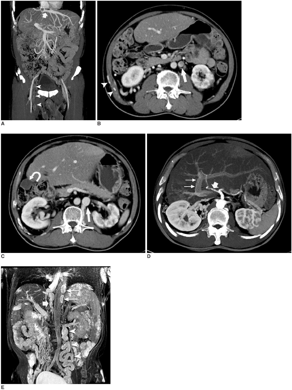
Fig. 1 62-years-old patient with right hepatic lobe agenesis, combined with deep venous thrombosis caused by congenital absence of inferior vena cava. A. Coronal thick (30 mm) slab maximum intensity projection CT image showed thrombus (arrowheads) occupying entire lumen of right femoral and external iliac vein. Left hepatic vein (arrow) drains into inferior vena cava (asterisk). B. Transverse CT scan obtained inferior to kidney shows missing IVC. Enlarged left testicular vein (arrow) and prominent anterior abdominal wall collateral veins (arrowheads) are present. C. Transverse contrast-enhanced CT scan obtained at hilus level of left kidney shows missing infrarenal segment belonging to IVC. Enlarged ascending lumbar vein and prominent left testicular vein (arrow) are present. Right hepatic lobe is absent and gallbladder (curved arrow) is identified in retrohepatic position. D. Transverse thick (30 mm) slab maximum intensity projection CT image of arterial phase shows left hepatic artery (large arrow) arising from celiac axis. Complete absence of right hepatic artery branch is noted, along with left portal vein (small arrows). E. Coronal MIP of two-dimensional time-of-flight MR image shows absence of infrarenal IVC, with enlarged azygos (large arrow) and hemiazygos vein (small arrows). Tortuous and enlarged left testicular vein (arrowheads) is also noted.
Reference
-
1. Bass JE, Redwine MD, Kramer LA, Huynh PT, Harris JH Jr. Spectrum of congenital anomalies of the inferior vena cava: cross-sectional imaging findings. Radiographics. 2000. 20:639–652.2. Bass JE, Redwine MD, Kramer LA, Harris JH Jr. Absence of the infrarenal inferior vena cava with preservation of the suprarenal segment as revealed by CT and MR venography. AJR Am J Roentgenol. 1999. 172:1610–1612.3. Gayer G, Luboshitz J, Hertz M, Zissin R, Thaler M, Lubetsky A, et al. Congenital anomalies of the inferior vena cava revealed on CT in patients with deep vein thrombosis. AJR Am J Roentgenol. 2003. 180:729–732.4. Chou CK, Mak CW, Lin MB, Tzeng WS, Chang JM. CT of agenesis and atrophy of the right hepatic lobe. Abdom Imaging. 1998. 23:603–607.5. Inoue T, Ito Y, Matsuzaki Y, Okauchi Y, Kondo H, Horiuchi N, et al. Hypogenesis of right hepatic lobe accompanied by portal hypertension: case report and review of 31 Japanese cases. J Gastroenterol. 1997. 32:836–842.6. d'Archambeau O, Verguts L, Myle J. Congenital absence of inferior vena cava. J Belge Radiol. 1990. 73:516–517.7. Gayer G, Apter S, Jonas T, Amitai M, Zissin R, Sella T, et al. Polysplenia syndrome detected in adulthood: report of eight cases and review of the literature. Abdom Imaging. 1999. 24:178–184.8. Gayer G, Zissin R, Strauss S, Hertz M. IVC anomalies and right renal aplasia detected on CT: a possible link? Abdom Imaging. 2003. 28:395–399.9. Radin DR, Colletti PM, Ralls PW, Boswell WD Jr, Halls JM. Agenesis of the right lobe of the liver. Radiology. 1987. 164:639–642.10. Zhang L, Yang G, Shen W, Qi J. Spectrum of the inferior vena cava: MDCT findings. Abdom Imaging. 2007. 32:495–503.
- Full Text Links
-
- Actions
-
Cited
- CITED
-
- Close
- Share
-
- Similar articles
-
- Absence of the Intrahepatic Inferior Vena Cava with Polysplenia Syndrome on Multidetector Computed Tomography: A Case Report
- Congenital Interruption of the Inferior Vena Cava with Azygos Continuation: A Case Report
- A Case of Congenital Absence of the Inferior Vena Cava
- Transposition of inferior vena cava
- Obstruction of the Hepatic Portion of the Inferior Vena Cava
