A "Benign" Sphenoid Ridge Meningioma Manifesting as a Subarachnoid Hemorrhage Associated with Tumor Invasion into the Middle Cerebral Artery
- Affiliations
-
- 1Department of Diagnostic Radiology, Ajou University School of Medicine, Gyeonggi-do, Korea. J978005@lycos.co.kr
- KMID: 1100095
- DOI: http://doi.org/10.3348/kjr.2008.9.s.s10
Abstract
- Meningioma rarely manifests as a subarachnoid hemorrhage (SAH), and invasion directly into a major intracranial artery is extremely rare. To the best of our knowledge, meningioma presenting with an SAH associated with major intracranial arterial invasion has never been reported. We present a case of sphenoid ridge meningotheliomatous meningioma manifesting as an SAH without pathologically atypical or malignant features, due to direct tumor invasion into the middle cerebral artery.
MeSH Terms
Figure
-
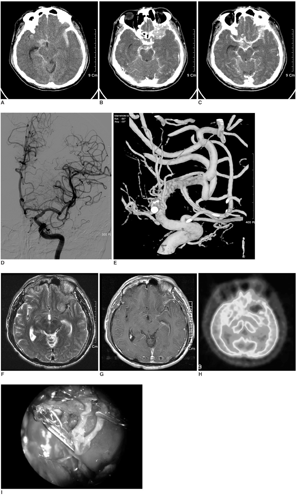
Fig. 1 Initial CT scans and angiographies of 53-year-old man that presented with sudden onset of severe headache. A. Noncontrast CT axial scan shows large amount of subarachnoid hemorrhage, especially in left sylvian fissure. B. Contrast-enhanced CT axial scan shows enhancing mass lesion around sphenoid ridge with combined bony destruction. C. Contrast-enhanced CT axial scan shows that mass is in contact with left middle cerebral artery. D. Digital subtraction angiography with selective injection of left internal carotid artery reveals no evidence of aneurysms or arteriovenous malformations. E. Three-dimensional rotational angiography demonstrates mild focal dilatation at proximal M2 portion of left middle cerebral artery. F. Axial T2-weighted MR image demonstrates hyperintense mass adjacent to left middle cerebral artery. G. Enhanced axial T1-weighted MR image demonstrates mass with mild enhancement. H. PET scan shows no definite uptake of mass lesion, suggesting benign or low-grade tumor. I. Intraoperative photomicrograph. Perforation at just distal portion of left middle cerebral artery bifurcation is noted.
Reference
-
1. Roser F, Nakamura M, Jacobs C, Vorkapic P, Samii M. Sphenoid wing meningiomas with osseous involvement. Surg Neurol. 2005. 64:37–43.2. Shaffrey ME, Dolenc VV, Lanzino G, Wolcott WP, Shaffrey CI. Invasion of the internal carotid artery by cavernous sinus meningiomas. Surg Neurol. 1999. 52:167–171.3. Kim DG, Park CK, Paek SH, Choe GY, Gwak HS, Yoo H, et al. Meningioma manifesting intracerebral hemorrhage: a possible mechanism of haemorrhage. Acta Neurochir (Wien). 2000. 142:165–168.4. Niiro M, Ishimaru K, Hirano H, Yunoue S, Kuratsu J. Clinico-pathological study of meningiomas with haemorrhagic onset. Acta Neurochir (Wien). 2003. 145:767–772.5. Bosnjak R, Derham C, Popovic M, Ravnik J. Spontaneous intracranial meningioma bleeding: clinicopathological features and outcome. J Neurosurg. 2005. 103:473–484.6. Locksley HB, Sahs AL, Sandler R. Report on the cooperative study of intracranial aneurysms and subarachnoid hemorrhage. 3. Subarachnoid hemorrhage unrelated to intracranial aneurysm and A-V malformation A study of associated diseases and prognosis. J Neurosurg. 1966. 24:1034–1056.7. Pluchino F, Lodrini S, Savoiardo M. Subarachnoid hemorrhage and meningioma. Report of two cases. Acta Neurochir (Wien). 1983. 68:45–53.8. Yucesoy K, Ozer H, Erdag N, Mertol T. Meningioma presented as subarachnoid haemorrhage: case report. Kobe J Med Sci. 1999. 45:213–219.9. Kotapka MJ, Kalia KK, Martinez AJ, Sekhar LN. Infiltration of the carotid artery by cavernous sinus meningioma. J Neurosurg. 1994. 81:252–255.
- Full Text Links
-
- Actions
-
Cited
- CITED
-
- Close
- Share
-
- Similar articles
-
- Sphenoid Ridge Meningioma Presenting as Acute Cerebral Infarction
- Subarachnoid Hemorrhage Due to a Ruptured Middle Cerebral Artery Bifurcation Aneurysm Superimposed by an Idiopathic Intracerebral Hematoma
- Traumatic Intracerebral and Subarachnoid Hemorrhage Due to a Ruptured Pseudoaneurysm of Middle Meningeal Artery Accompanied by a Medial Sphenoid Wing Dural Arteriovenous Fistula
- Roentgenologic analysis of meningioma
- The statistical observation for frequancy of occurence of the anencephalus
