Korean J Ophthalmol.
2006 Mar;20(1):26-32. 10.3341/kjo.2006.20.1.26.
Comparison of Retinal Nerve Fiber Layer Measurements between NTG and HTG using GDx-VCC
- Affiliations
-
- 1Department of Ophthalmology, University of Soonchunhyang, College of Medicine, Seoul, Korea.
- 2Department of Ophthalmology, University of Ulsan, College of Medicine, Asan Medical Center, Seoul, Korea. mskook@amc.seoul.kr
- KMID: 1099069
- DOI: http://doi.org/10.3341/kjo.2006.20.1.26
Abstract
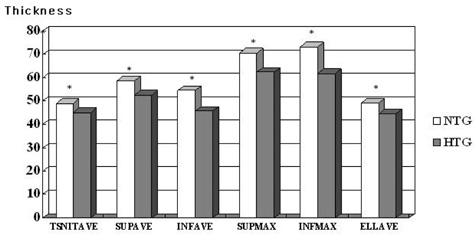
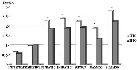
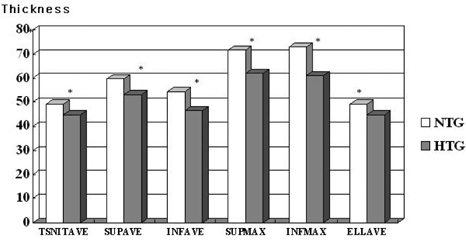
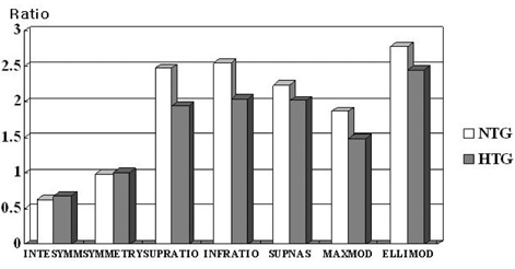
- PURPOSE: To compare quantitative polarimetric measurements in eyes with NTG and HTG using GDx-VCC. Both groups were matched by age and glaucoma stage based on the Humphrey visual field test. METHODS: We retrospectively reviewed the records of 146 patients who underwent Humphrey field analysis (HFA) and GDx-VCC. We compared outcomes of retinal nerve fiber layer (RNFL) parameters among the three groups by ANOVA and between each pair of groups using the Tukey-Kramer Post-Hoc test. We also evaluated the sensitivity and specificity of GDx-VCC in detecting glaucoma in each group. RESULTS: The mean age and HFA mean deviation (MD) were 55.6+/-9.5 years and -0.8+/-1.5 dB in 47 control patients, 59.4+/-9.0 years and -5.77+/-4.38 dB in 49 NTG patients, and 59.4+/-11.7 years and -8.09+/-6.77 dB in 51 HTG patients, respectively. All thickness parameters were lower in HTG patients compared to NTG patients, but there were no significant differences in ratio parameters between age-matched early HTG and NTG patients. The sensitivity of GDx-VCC was significantly higher in both early and total HTG patients compared to the respective groups of NTG patients. CONCLUSIONS: Compared to eyes with NTG, eyes with HTG showed reduced RNFL thickness and ratio parameters when patients were age and visual field matched. GDx-VCC appeared to be more sensitive in detecting RNFL damage in HTG patients.
Keyword
MeSH Terms
Figure
Reference
-
1. Yamazaki Y, Koide C, Miyazawa T, et al. Comparison of the retinal nerve fiber layer in high and normal tension glaucoma. Graefes Arch Clin Exp Ophthalmol. 1991. 229:517–520.2. Spaeth GL. Low tension glaucoma: its diagnosis and management. Doc Ophthalmol Proc Ser. 1980. 22:263–287.3. Flammer J. Psychophysics in glaucoma. A modified concept of the disease. Doc Ophthalmol Proc Ser. 1985. 43:11–17.4. Kubota T, Khalil AK, Honda M, et al. Comparative study of retinal nerve fiber layer damage in Japanese patients with normal and high tension glaucoma. J Glaucoma. 1999. 8:363–366.5. Iester M, Mikelberg FS. Optic nerve head morphologic characteristics in high tenision and normal tension glaucoma. Arch Ophthalmol. 1999. 117:1010–1013.6. Kook MS, Lee SU, Sung KR, et al. Pattern of retinal nerve fiber layer damage in Korean eyes with normal-tension glaucoma and hemifield visual field defect. Graefes Arch Clin Exp Ophthalmol. 2002. 240:448–456.7. Zangwill L, Berry CA, Garden VS, Weinreb RN. Reproducibility of retardation measurements with the nerve fiber analyzer II. J Glaucoma. 1997. 6:384–389.8. Hoh ST, Ishikawa H, Greenfield DS, et al. Peripapillary nerve fiber layer thickness measurement reproducibility using scanning laser polarimetry. J Glaucoma. 1998. 7:12–15.9. Weinreb RN, Shakiba S, Zangwill L. Scanning laser polarimery to measure the nerve fiber layer of normal and glaucomatous eyes. Am J Ophthalmol. 1995. 119:627–636.10. Knighton RW, Huang XR. Linear birefringence of the central human cornea. Invest Ophthalmol Vis Sci. 2002. 43:82–86.11. Greenfield DS, Knighton RW, Feuer WJ, et al. Correction for corneal polarization axis improves discriminating power of scanning laser polarimetry. Am J Ophthalmol. 2002. 134:27–33.12. Zhou Q, Weinreb RN. Individualized compensation of anterior segment birefringence during scanning laser polarimetry. Invest Ophthalmol Vis Sci. 2002. 43:2221–2228.13. Greenfield DS, Knighton RW, Huang XR. Effect of corneal polarization axis on assessment of retinal nerve fiber layer thickness by scanning laser polarimetry. Am J Ophthalmol. 2000. 129:715–722.14. Knighton W, Huang XR, Greenfield DS. Analytical model of scanning laser polarimetry for retinal nerve fiber layer assessment. Invest Ophthalmol Vis Sci. 2002. 43:383–392.15. Weinreb RN, Bowd C, Greenfield DS, Zangwill LM. Measurement of the magnitude and axis of corneal polarization with scanning laser polarimetry. Arch Ophthalmol. 2002. 120:901–906.16. Choplin NT, Zhou Q, Knighton RW. Effect of individualized compensation for anterior segment birefringence on retinal nerve fiber layer assessments as determined by scanning laser polarimetry. Ophthalmology. 2003. 110:719–725.17. Weinreb RN, Bowd C, Zangwill LM. Glaucoma detection using scanning laser polarimetry with variable corneal polarization compensation. Arch Ophthalmol. 2003. 121:218–224.18. Matsumoto C, Shirato S, Haneda M, et al. Study of retinal nerve fiber layer thickness within normal hemivisual field in primary open angle glaucoma and normal-tension glaucoma. Jpn J Ophthalmol. 2003. 47:22–27.19. Kook MS, Sung K, Kim S, et al. Study of retinal nerve fiber layer thickness in eyes with high tension glaucoma and hemifield defect. Br J Ophthalmol. 2001. 85:1167–1170.20. Medeiros FA, Zangwill LM, Bowd C, Weinreb RN. Comparison of the GDx VCC scanning laser polarimeter, HRT II confocal scanning laser ophthalmoscope, and stratus OCT optical coherence tomograph for the detection of glaucoma. Arch Ophthalmol. 2004. 122:827–837.21. Funaki S, Shirakashi M, Abe H. Relation between size of optic disc and thickness of retinal nerve fiber layer in normal subjects. Br J Ophthalmol. 1998. 82:1242–1245.
- Full Text Links
- Actions
-
Cited
- CITED
-
- Close
- Share
- Similar articles
-
- Longitudinal Analysis of Retinal Nerve Fiber Layer Thickness With GDx-VCC in Glaucoma Suspect
- The Relationship between Optical Coherence Tomography and Scanning Laser Polarimetry Measurements in Glaucoma
- Scanning Laser Polarimetry and Optical Coherence Tomography for Detection of Retinal Nerve Fiber Layer Defects
- Normal Retinal Nerve Fiber Layer Thickness in the Peripapillary Region of Koreans Measured by Scanning Laser Polarimeter
- Comparison of OCT and HRT Findings Among Normal, Normal Tension Glaucoma, and High Tension Glaucoma


