Korean J Ophthalmol.
2011 Oct;25(5):362-365. 10.3341/kjo.2011.25.5.362.
A Case of Cholesterosis Bulbi with Secondary Glaucoma Treated by Vitrectomy and Intravitreal Bevacizumab
- Affiliations
-
- 1Department of Ophthalmology, Eulji General Hospital, Eulji University School of Medicine, Seoul, Korea. pjs4106@eulji.ac.kr
- 2Department of Laboratory Medicine, Eulji General Hospital, Eulji University School of Medicine, Seoul, Korea.
- KMID: 1098643
- DOI: http://doi.org/10.3341/kjo.2011.25.5.362
Abstract
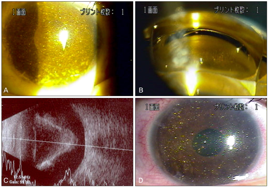
- We report on a case of cholesterosis bulbi concurrent with secondary glaucoma. A 36-year-old man, with a history of long-standing retinal detachment in his right eye after the irrigation and aspiration of a congenital cataract, presented with a clinical picture of elevated intraocular pressure and ocular pain. Upon slit-lamp examination, we found a ciliary injection and a pseudohypopyon of polychromatic crystals. Gonioscopic examination revealed a large amount of crystals deposited on the trabecular meshwork and mild rubeosis iridis, but the neovascularization of the angle could not be clearly confirmed due to the presence of so many crystals. Pars plana vitrectomy was performed to remove clusters of crystals and bevacizumab was injected intravitreally to treat iris neovascularization. Aqueous aspirate was examined by light microscopy and the typical highly refringent cholesterol crystals were identified. Intraocular pressure returned to a normal level after the bevacizumab injection, although severe cholesterosis was still evident in the anterior chamber. To our knowledge, this would be the first Korean case of cholesterosis bulbi combined with chronic retinal detachment and presumed neovascular glaucoma, which was treated by pars plana vitrectomy and intravitreal bevacizumab injection.
MeSH Terms
-
Adult
Angiogenesis Inhibitors/therapeutic use
Anterior Chamber/*metabolism
Antibodies, Monoclonal, Humanized/*therapeutic use
*Cholesterol
Eye Diseases/complications/metabolism
Follow-Up Studies
Glaucoma/surgery
Glaucoma, Neovascular/drug therapy/*etiology/surgery
Humans
Intraocular Pressure
Male
Vitrectomy/*methods
Figure
Reference
-
1. Mielke J, Freudenthaler N, Schlote T, Bartz-Schmidt KU. Pseudohypopyon of cholesterol crystals occurring 16 years after retinal detachment in x-linked retinoschisis. Klin Monbl Augenheilkd. 2001. 218:741–743.2. Wand M, Smith TR, Cogan DG. Cholesterosis bulbi: the ocular abnormality known as synchysis scintillans. Am J Ophthalmol. 1975. 80:177–183.3. Forsius H. Cholesterol-crystals in the anterior chamber. A clinical and chemical study of 7 cases. Acta Ophthalmol (Copenh). 1961. 39:284–301.4. Shields JA, Eagle RC Jr, Fammartino J, et al. Coats' disease as a cause of anterior chamber cholesterolosis. Arch Ophthalmol. 1995. 113:975–977.5. Eagle RC Jr, Yanoff M. Cholesterolosis of the anterior chamber. Albrecht Von Graefes Arch Klin Exp Ophthalmol. 1975. 193:121–134.6. Kumar S. Cholesterol crystals in the anterior chamber. Br J Ophthalmol. 1963. 47:295–299.7. Spraul CW, Grossniklaus HE. Vitreous hemorrhage. Surv Ophthalmol. 1997. 42:3–39.8. Andrews JS, Lynn C, Scobey JW, Elliott JH. Cholesterosis bulbi. Case report with modern chemical identification of the ubiquitous crystals. Br J Ophthalmol. 1973. 57:838–844.9. Kennedy CJ. The pathogenesis of polychromatic cholesterol crystals in the anterior chamber. Aust N Z J Ophthalmol. 1996. 24:267–273.10. Eibschitz-Tsimhoni M, Johnson MW, Johnson TM, Moroi SE. Coats' syndrome as a cause of secondary open-angle glaucoma. Ophthalmic Surg Lasers Imaging. 2003. 34:312–314.11. Gruber E. Crystals in the anterior chamber. Am J Ophthalmol. 1955. 40:817–827.12. Hubbersty FS, Gourlay JS. Secondary glaucoma due to spontaneous rupture of the lens capsule. Br J Ophthalmol. 1953. 37:432–435.13. Yu YS, Kwak HW, Youn DH. Cholesterol crystals in the anterior chamber. J Korean Ophthalmol Soc. 1980. 21:117–119.14. Shyn KW, Koo JY, Lee YH. Two cases of cholesterosis bulbi. J Korean Ophthalmol Soc. 1986. 27:99–103.
- Full Text Links
- Actions
-
Cited
- CITED
-
- Close
- Share
- Similar articles
-
- A Case of Retinal Hemorrhage Following a Dexamethasone Intravitreal Implant
- Intravitreal Bevacizumab and Subsequent Trabeculectomy with Mitomycin C for Neovascular Glaucoma with Previous Sutureless Vitrectomy
- A Case of Intravitreal Cysticercosis with Neovascular Glaucoma
- Intravitreal Bevacizumab Injection as Preoperative Adjuvant of Vitrectomy for Proliferative Diabetic Retinopathy
- Trabeculectomy Combined with Pars Plana Vitrectomy


