Korean J Radiol.
2009 Oct;10(5):464-471. 10.3348/kjr.2009.10.5.464.
Comparative Analysis of Logistic Regression, Support Vector Machine and Artificial Neural Network for the Differential Diagnosis of Benign and Malignant Solid Breast Tumors by the Use of Three-Dimensional Power Doppler Imaging
- Affiliations
-
- 1Department of Surgery, Changhua Christian Hospital, Changhua, Taiwan. 115045@cch.org.tw
- 2Department of Research, Changhua Christian Hospital, Changhua, Taiwan.
- 3Department of Computer Science and Information Engineering, Tunghai University, Taichung, Taiwan.
- 4Comprehensive Breast Cancer Center, Changhua Christian Hospital, Changhua, Taiwan.
- 5Department of Medical Imaging, Changhua Christian Hospital, Changhua, Taiwan.
- KMID: 1093961
- DOI: http://doi.org/10.3348/kjr.2009.10.5.464
Abstract
OBJECTIVE
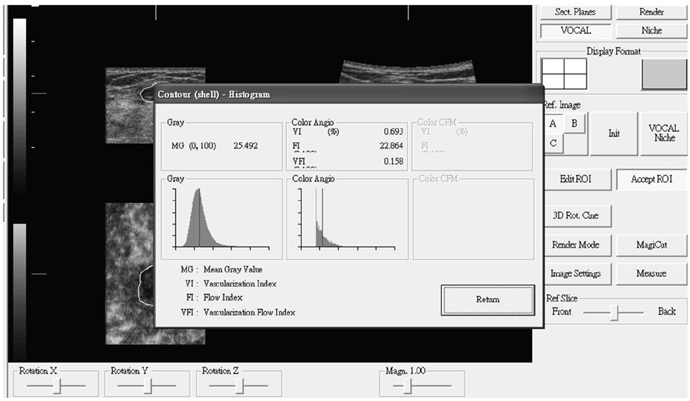
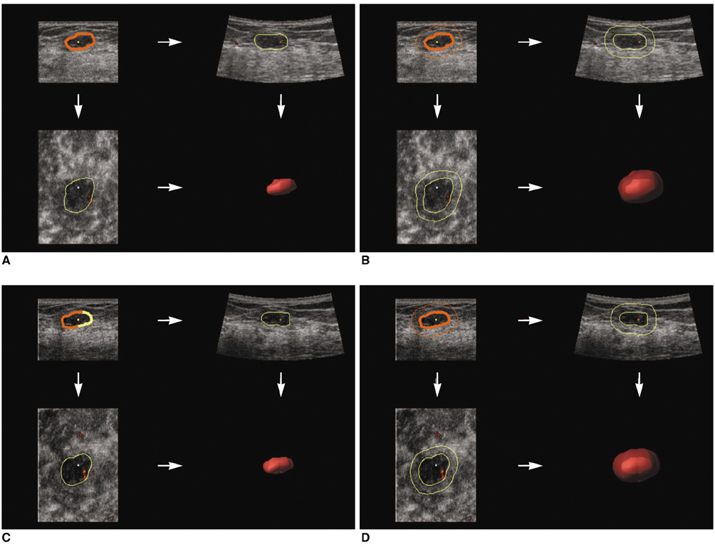
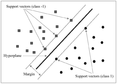
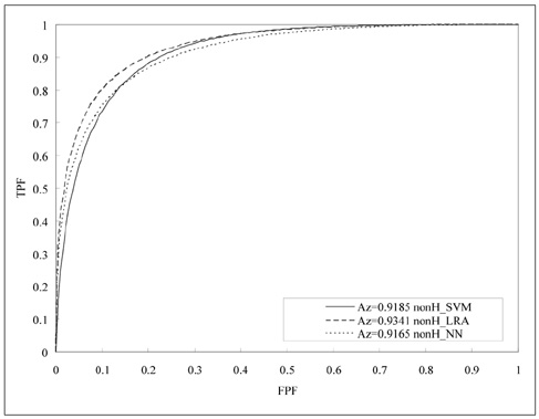
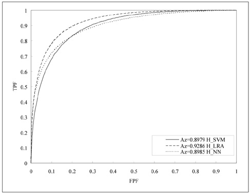
Logistic regression analysis (LRA), Support Vector Machine (SVM) and a neural network (NN) are commonly used statistical models in computer-aided diagnostic (CAD) systems for breast ultrasonography (US). The aim of this study was to clarify the diagnostic ability of the use of these statistical models for future applications of CAD systems, such as three-dimensional (3D) power Doppler imaging, vascularity evaluation and the differentiation of a solid mass. MATERIALS AND METHODS: A database that contained 3D power Doppler imaging pairs of non-harmonic and tissue harmonic images for 97 benign and 86 malignant solid tumors was utilized. The virtual organ computer-aided analysis-imaging program was used to analyze the stored volumes of the 183 solid breast tumors. LRA, an SVM and NN were employed in comparative analyses for the characterization of benign and malignant solid breast masses from the database. RESULTS: The values of area under receiver operating characteristic (ROC) curve, referred to as Az values for the use of non-harmonic 3D power Doppler US with LRA, SVM and NN were 0.9341, 0.9185 and 0.9086, respectively. The Az values for the use of harmonic 3D power Doppler US with LRA, SVM and NN were 0.9286, 0.8979 and 0.9009, respectively. The Az values of six ROC curves for the use of LRA, SVM and NN for non-harmonic or harmonic 3D power Doppler imaging were similar. CONCLUSION: The diagnostic performances of these three models (LRA, SVM and NN) are not different as demonstrated by ROC curve analysis. Depending on user emphasis for the use of ROC curve findings, the use of LRA appears to provide better sensitivity as compared to the other statistical models.
Keyword
MeSH Terms
-
Adolescent
Adult
Aged
Aged, 80 and over
*Artificial Intelligence
Breast Neoplasms/*ultrasonography
Diagnosis, Computer-Assisted
Diagnosis, Differential
Female
Humans
Image Interpretation, Computer-Assisted
Imaging, Three-Dimensional/*statistics & numerical data
Logistic Models
Middle Aged
*Neural Networks (Computer)
Predictive Value of Tests
ROC Curve
Sensitivity and Specificity
Ultrasonography, Doppler/*statistics & numerical data
Ultrasonography, Mammary/*statistics & numerical data
Figure
Reference
-
1. Cho N, Moon WK, Park JS, Cha JH, Jang M, Seong MH. Nonpalpable breast masses: evaluation by US elastography. Korean J Radiol. 2008. 9:111–118.2. Chen DR, Chang RF, Chen WM, Moon WK. Computer-aided diagnosis for 3-dimensional breast ultrasonography. Arch Surg. 2003. 138:296–302.3. Kuo SJ, Hsiao YH, Huang YL, Chen DR. Classification of benign and malignant breast tumors using neural networks and three-dimensional power Doppler ultrasound. Ultrasound Obstet Gynecol. 2008. 32:97–102.4. Rosen EL, Soo MS. Tissue harmonic imaging sonography of breast lesions: improved margin analysis, conspicuity, and image quality compared to conventional ultrasound. Clin Imaging. 2001. 25:379–384.5. Szopinski KT, Pajk AM, Wysocki M, Amy D, Szopinska M, Jakubowski W. Tissue harmonic imaging: utility in breast sonography. J Ultrasound Med. 2003. 22:479–487.6. Seo BK, Oh YW, Kim HR, Kim HW, Kang CH, Lee NJ, et al. Sonographic evaluation of breast nodules: comparison of conventional, real-time compound, and pulse-inversion harmonic images. Korean J Radiol. 2002. 3:38–44.7. Hsiao YH, Kuo SJ, Liang WM, Huang YL, Chen DR. Intra-tumor flow index can predict the malignant potential of breast tumor: dependent on age and volume. Ultrasound Med Biol. 2008. 34:88–95.8. Chang RF, Huang SF, Moon WK, Lee YH, Chen DR. Solid breast masses: neural network analysis of vascular features at three-dimensional power Doppler US for benign or malignant classification. Radiology. 2007. 243:56–62.9. Chen DR, Chang RF, Huang YL. Computer-aided diagnosis applied to US of solid breast nodules by using neural networks. Radiology. 1999. 213:407–412.10. Chen DR, Chang RF, Huang YL, Chou YH, Tiu CM, Tsai PP. Texture analysis of breast tumors on sonograms. Semin Ultrasound CT MR. 2000. 21:308–316.11. Chen DR, Chang RF, Kuo WJ, Chen MC, Huang YL. Diagnosis of breast tumors with sonographic texture analysis using wavelet transform and neural networks. Ultrasound Med Biol. 2002. 28:1301–1310.12. Seker H, Odetayo MO, Petrovic D, Naguib RN, Bartoli C, Alasio L, et al. Assessment of nodal involvement and survival analysis in breast cancer patients using image cytometric data: statistical, neural network and fuzzy approaches. Anticancer Res. 2002. 22:433–438.13. Tourassi GD. Journey toward computer-aided diagnosis: role of image texture analysis. Radiology. 1999. 213:317–320.14. Joo S, Yang YS, Moon WK, Kim HC. Computer-aided diagnosis of solid breast nodules: use of an artificial neural network based on multiple sonographic features. IEEE Trans Med Imaging. 2004. 23:1292–1300.15. Chen CM, Chou YH, Han KC, Hung GS, Tiu CM, Chiou HJ, et al. Breast lesions on sonograms: computer-aided diagnosis with nearly setting-independent features and artificial neural networks. Radiology. 2003. 226:504–514.16. Chang RF, Wu WJ, Moon WK, Chou YH, Chen DR. Support vector machines for diagnosis of breast tumors on US images. Acad Radiol. 2003. 10:189–197.17. Huang YL, Chen DR. Support vector machines in sonography: application to decision making in the diagnosis of breast cancer. Clin Imaging. 2005. 29:179–184.18. Zhang J, Wang Y, Dong Y, Wang Y. Ultrasonographic feature selection and pattern classification for cervical lymph nodes using support vector machines. Comput Methods Programs Biomed. 2007. 88:75–84.19. Jarvela IY, Sladkevicius P, Kelly S, Ojha K, Nargund G, Campbell S. Three-dimensional sonographic and power Doppler characterization of ovaries in late follicular phase. Ultrasound Obstet Gynecol. 2002. 20:281–285.20. Chang CH, Yu CH, Ko HC, Chen CL, Chang FM. Three-dimensional power Doppler ultrasound for the assessment of the fetal brain blood flow in normal gestation. Ultrasound Med Biol. 2003. 29:1273–1279.21. Alcazar JL. Tumor angiogenesis assessed by three-dimensional power Doppler ultrasound in early, advanced and metastatic ovarian cancer: a preliminary study. Ultrasound Obstet Gynecol. 2006. 28:325–329.22. Chang RF, Wu WJ, Moon WK, Chen DR. Improvement in breast tumor discrimination by support vector machines and speckle-emphasis texture analysis. Ultrasound Med Biol. 2003. 29:679–686.23. Song JH, Venkatesh SS, Conant EA, Arger PH, Sehgal CM. Comparative analysis of logistic regression and artificial neural network for computer-aided diagnosis of breast masses. Acad Radiol. 2005. 12:487–495.
- Full Text Links
- Actions
-
Cited
- CITED
-
- Close
- Share
- Similar articles
-
- Machine Learning on Early Diagnosis of Depression
- Solid Breast Lesions: Evaluation with Power versus Conventional Color Doppler Sonography
- Resistive Index in Breast Tumors: Usefulness on Differentiation between Benign and Malignant Lesions
- Anesthesia research in the artificial intelligence era
- A Narrative Review on the Application of Artificial Intelligence on the Diagnosis and Outcome Prediction for Spinal Diseases

