Generalized Lymphangiomatosis: Radiologic Findings in Three Pediatric Patients
- Affiliations
-
- 1Department of Radiology, University of Ulsan College of Medicine, Asan Medical Center, Seoul, Korea. hwgoo@amc.seoul.kr
- KMID: 1092553
- DOI: http://doi.org/10.3348/kjr.2006.7.4.287
Abstract
- Generalized lymphangiomatosis is a rare disease that is characterized by widespread bony and soft tissue involvement of lymphangioma. Radiological evaluation is crucial because the site and extent of the lymphangioma are important prognostic factors. We reported here on three cases of generalized lymphangiomatosis and all three cases showed similar radiologic findings, but a different clinical course. The CT, US and MR images showed sharply defined, non-enhanced cystic lesions involving the mediastinum, bones, spleen, lung and lower neck. The whole body MR imaging with the short tau inversion recovery (STIR) sequence showed good capability for evaluating the extent of disease.
MeSH Terms
Figure
-
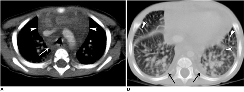
Fig. 1 Case 1. A 4-year-old boy. A. Contrast enhanced CT shows a mediastinal mass (arrowheads) enveloping the thoracic great vessels and thymus. The mass extends around the trachea and esophagus (arrow). B. The CT image with the lung window setting shows interstitial thickenings (arrowheads) and bilateral pleural effusions (arrows).
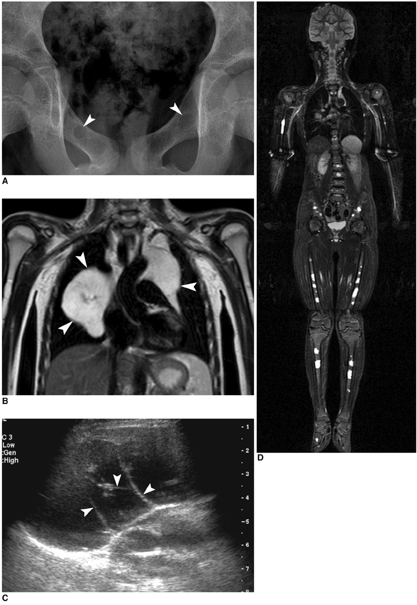
Fig. 2 Case 2. An 8-year-old boy. A. Pelvic bone radiograph shows well-defined osteolytic lesions (arrowheads) with thin sclerotic rims in both pubic bones. B. The coronal T2 weighted MR image (TR/TE = 1200/90, flip angle = 90°, slice thickness = 6 mm) shows a homogenous high-signal intensity mass in the mediastinum (arrowheads). C. On ultrasound, the mass is delineated as a multiseptated anechoic mass. Note the multiple septa in the mass (arrowheads). D. The coronal short tau inversion recovery whole body MR image shows multiple high signal intensity lesions involving vertebrae, pelvic bones, both humeri, both femurs, both tibias, the ribs, and calvarium.
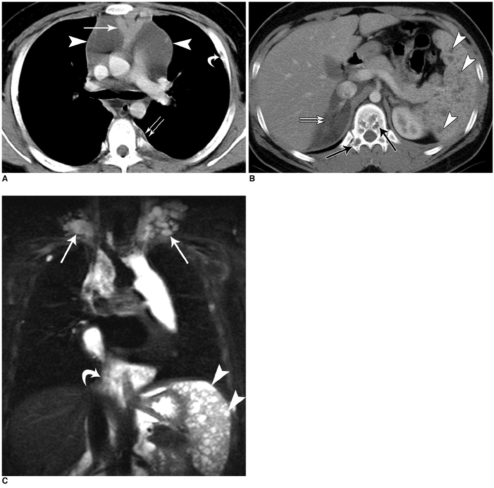
Fig. 3 Case 3. An 11-year-old boy. A. The contrast enhanced CT shows a mediastinal mass (arrowheads) enveloping the thoracic great vessels and thymus (arrow). The mass extends to the posterior mediastinum (double arrow). Note the well-defined osteolytic lesion in the left fourth rib (curved arrow). B. Contrast enhanced CT shows multiple low-density lesions (arrowheads) in the spleen. Multiple well-defined osteolytic lesions with sclerotic rims are noted in the thoracic vertebra (arrows). Note the mass extending to the retroperitoneum (double arrow). C. The coronal short tau inversion recovery MR image shows multiple high-signal intensity lesions involving the bilateral lower neck (arrows), the mediastinum and the spleen (arrowheads). Note the mass extending to the posterior mediastinum (curved arrow).
Cited by 1 articles
-
A Case of a Retroperitoneal Cystic Lymphangioma Treated by Percutaneous Catheter Drainage and Sclerotherapy
Hyun Sik Kang, Seung Hyung Kim, Bong Soo Kim, Ki Soo Kang
Korean J Pediatr Gastroenterol Nutr. 2010;13(1):86-91. doi: 10.5223/kjpgn.2010.13.1.86.
Reference
-
1. Brown LR, Reiman HM, Rosenow EC 3rd, Gloviczki PM, Divertie MB. Intrathoracic lymphangioma. Mayo Clin Proc. 1986. 61:882–892.2. Levey DS, MacCormack LM, Sartoris DJ, Haghighi P, Resnick D, Thorne R. Cystic angiomatosis: case report and review of the literature. Skeletal Radiol. 1996. 25:287–293.3. Wunderbaldinger P, Paya K, Partik B, Turetschek K, Hormann M, Horcher E, et al. CT and MR imaging of generalized cystic lymphangiomatosis in pediatric patients. AJR Am J Roentgenol. 2000. 174:827–832.4. Singh S, Baboo ML, Pathak IC. Cystic lymphangioma in children: report of 32 children including lesions at rare sites. Surgery. 1971. 69:947–951.5. Shah AR, Dinwiddie R, Woolf D, Ramani R, Higgins JN, Matthew DJ. Generalized lymphangiomatosis and chylothorax in the pediatric age group. Pediatr Pulmonol. 1992. 12:126–130.6. Canady AI, Chou SN. Cervical lymphangiomatosis with progressive craniospinal deformity. Neurosurgery. 1980. 6:422–425.7. Laffan EE, O'Connor R, Ryan SP, Donoghue VB. Whole-body magnetic resonance imaging: a useful additional sequence in paediatric imaging. Pediatr Radiol. 2004. 34:472–480.8. Azouz EM, Saigal G, Rodriguez MM, Podda A. Langerhans' cell histiocytosis: pathology, imaging and treatment of skeletal involvement. Pediatr Radiol. 2005. 35:103–115.9. Scafidi DE, McLeary MS, Young LW. Diffuse neonatal gastrointestinal hemangiomatosis: CT findings. Pediatr Radiol. 1998. 28:512–514.10. Rostom AY. Treatment of thoracic lymphangiomatosis. Arch Dis Child. 2000. 83:138–139.
- Full Text Links
-
- Actions
-
Cited
- CITED
-
- Close
- Share
-
- Similar articles
-
- Generalized Lymphangiomatosis: A Case Report
- A Case of Disseminated Lymphangiomatosis Involving Mediastinum, Bone, Spleen and Retroperitoneum in an Asymptomatic Healthy Child
- Metachronous Bilateral Renal Lymphangiomatosis Mimicking as a Simple Renal Cyst
- Colonic Lymphangiomatosis with Normal Colonoscopic Finding in an Adult
- Lymphangiomatosis of Bone and Soft Tissue: A Case Report
