J Vet Sci.
2007 Sep;8(3):229-236. 10.4142/jvs.2007.8.3.229.
Retrospective study of canine cutaneous tumors in Korea
- Affiliations
-
- 1Department of Veterinary Pathology, College of Veterinary Medicine, Seoul National University, Seoul 151-742, Korea. daeyong@snu.ac.kr
- 2Department of Veterinary Medicine, Cheju National University, Jeju 690-756, Korea
- 3School of Veterinary Medicine, Kangwon National University, Chuncheon 200-701, Korea
- 4Department of Laboratory Animal Medicine, College of Veterinary Medicine, Konkuk University, Seoul 143-701, Korea
- KMID: 1090796
- DOI: http://doi.org/10.4142/jvs.2007.8.3.229
Abstract
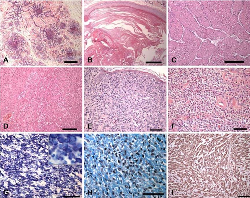
- Over the 42 month period from January 2003 to June2006, a total of 2,952 canine biopsy specimens werereceived from the Veterinary Medical Teaching Hospitalof Seoul National University and from veterinary practitionersacross the nation. Out of these, 748 (25.34%) cases werediagnosed as canine cutaneous tumors in the Departmentof Veterinary Pathology, College of Veterinary Medicine,Seoul National University, Korea. Thirty-eight differenttypes of cutaneous tumors were identified and categorizedinto epithelial and melanocytic tumors (56.95%), mesenchymaltumors (38.90%), and hematopoietic tumors (4.14%)located in the skin. Among these, 69.25% were benign and30.74% were malignant. The top ten most frequentlydiagnosed cutaneous tumors were epidermal and follicularcysts (12.70%), lipoma (11.36%), mast cell tumors (8.82%),cutaneous histiocytoma (7.49%), basal cell tumors (6.82%),sebaceous gland adenoma (6.68%), sebaceous glandhyperplasia (5.08%), hepatoid gland adenoma (3.61%),apocrine adenocarcinoma (3.07%), and fibroma (2.81%),in order of prevalence. They comprised 68.45% of allcutaneous tumors. These top ten cutaneous tumors weredistributed on the trunk (30.08%), head and neck(20.9%), extremities (19.14%), anal and perianal area(8.59%), and tail (3.91%). The age of the dogs with the tenmost frequent tumors had a mean age of 8.3 years, with arange of 2 months to 19 years. When all types of tumorswere considered together in the entire population, therewas no difference in incidence according to sex.
Keyword
MeSH Terms
Figure
Cited by 1 articles
-
Clinical Outcomes of Surgically Managed Spontaneous Tumors in 114 Client-owned Dogs
Ji-Won Choi, Hun-Young Yoon, Soon-Wuk Jeong
Immune Netw. 2016;16(2):116-125. doi: 10.4110/in.2016.16.2.116.
Reference
-
1. Bostock DE. Neoplasms of the skin and subcutaneous tissues in dogs and cats. Br Vet J. 1986. 142:1–19.
Article2. Brodey RS. Canine and feline neoplasia. Adv Vet Sci Comp Med. 1970. 14:309–354.3. Finnie JW, Bostock DE. Skin neoplasia in dogs. Aust Vet J. 1979. 55:602–604.
Article4. Fox LE. Morrison WB, editor. Mast cell tumors. Cancer in Dogs and Cats: Medical and Surgical Management. 2002. 2nd ed. Jackson: Teton NewMedia;451–468.5. Goldschmidt MH, Hendrick MJ. Meuten DJ, editor. Tumors of the skin and soft tissues. Tumors in Domestic Animals. 2002. 4th ed. Ames: Iowa State Press;45–117.
Article6. Goldschmidt MH, Shofer FS. Skin Tumors of the Dog and Cat. 1992. New York: Pergamon Press;1–295.7. Gross TL, Ihrke PJ, Walder EJ, Affolter VK, editors. Skin Diseases of the Dog and Cat Clinical and Histopathologic Diagnosis. 2005. 2nd ed. Ames: Blackwell Science;561–882.8. Kaldrymidou H, Leontides L, Koutinas AF, Saridomichelakis MN, Karayannopoulou M. Prevalence, distribution and factors associated with the presence and the potential for malignancy of cutaneous neoplasms in 174 dogs admitted to a clinic in northern Greece. J Vet Med A Physiol Pathol Clin Med. 2002. 49:87–91.
Article9. Keller ET, Madewell BR. Location and types of neoplasms in immature dogs: 69 cases (1964-1989). J Am Vet Med Assoc. 1992. 200:1530–1532.10. Ladds PW, Kraft H, Sokale A, Trueman KF. Neoplasms of the skin of dogs in tropical Queensland. Aust Vet J. 1983. 60:87–88.
Article11. Rothwell TLW, Howlett CR, Middleton DJ, Griffiths DA, Duff BC. Skin neoplasms of dogs in Sydney. Aust Vet J. 1987. 64:161–164.
Article12. Sanja AK, Kukolj V, Marinković D, Milijana K. Retrospective study of canine epithelial and melanocytic tumors. Acta Vet (Beograd). 2005. 55:319–326.
Article13. Schultheiss PC. Histologic features and clinical outcomes of melanomas of lip, haired skin, and nail bed locations of dogs. J Vet Diagn Invest. 2006. 18:422–425.
Article14. Scott DW, Miller WH, Griffin CE. Muller & Kirk's Small Animal Dermatology. 2001. 6th ed. Philadelphia: Saunders;1236–1414.15. Strafuss AC. Skin tumors. Vet Clin North Am Small Anim Pract. 1985. 15:473–492.
Article16. Thomas RC, Fox LE. Morrison WB, editor. Tumors of the skin and subcutis. Cancer in Dogs and Cats: Medical and Surgical Management. 2002. 2nd ed. Jackson: Teton NewMedia;469–488.17. Vail DM, Withrow SJ. Withrow SJ, MacEwen EG, editors. Tumors of the skin and subcutaneous tissues. Small Animal Clinical Oncology. 2001. 3rd ed. Philadelphia: Saunders;233–260.
Article
- Full Text Links
- Actions
-
Cited
- CITED
-
- Close
- Share
- Similar articles
-
- A Statistical Study of Cutaneous Malignant Tumors and Premalignant Lesions in Southeastern Gyeonggi-do Province over an 11-year Period (2006~2016)
- A Clinical Investigation of Cutaneous Malignant Tumors in Western Gyeongnam Province over 11 Years
- A Clinical Study of Cutaneous Tumors of the Head and Neck
- Alpha basic crystallin expression in canine mammary tumors
- Assessment of prognostic factors in dogs with mammary gland tumors: 60 cases (2014-2020)


