Diagnostic Performance of CT Colonography for the Detection of Colorectal Polyps
- Affiliations
-
- 1Department of Radiology, Song-Do Hospital, Seoul, Korea. bugbearsyun@naver.com
- 2Department of Gastroenterology, East-West Neo Medical Center,Kyung Hee University, Seoul, Korea.
- 3Department of Colorectal Surgery, Song-Do Hospital, Seoul, Korea.
- KMID: 1089435
- DOI: http://doi.org/10.3348/kjr.2007.8.6.484
Abstract
- OBJECTIVE: To investigate the diagnostic value of CT colonography for the detection of colorectal polyps. MATERIALS AND METHODS: From December 2004 to December 2005, 399 patients underwent CT colonography and follow-up conventional colonoscopy. We excluded cases of advanced colorectal cancer. We retrospectively analyzed the CT colonography findings and follow-up conventional colonoscopy findings of 113 patients who had polyps more than 6 mm in diameter. Radiologists using 3D and 2D computer generated displays interpreted the CT colonography images. The colonoscopists were aware of the CT colonography findings before the procedure. RESULTS: CT colonography detected 132 polyps in 107 of the 113 patients and conventional colonoscopy detected 114 colorectal polyps more than 6 mm in diameter in 87 of the 113 patients. The sensitivity of CT colonography analyzed per polyp was 91% (41/45) for polyps more than 10 mm in diameter and 89% (101/114) for polyps more than 6 mm in diameter. Thirteen polyps were missed by CT colonography and were detected on follow-up conventional colonoscopy. CONCLUSION: CT colonography is a sensitive diagnostic tool for the detection of colorectal polyps and adequate bowel preparation, optimal bowel distention and clinical experience are needed to reduce the rate of missing appropriate lesions.
MeSH Terms
-
Adult
Aged
Aged, 80 and over
Colonic Polyps/*diagnosis
Colonography, Computed Tomographic/*methods
Colonoscopy/methods
Colorectal Neoplasms/*diagnosis
Contrast Media/administration & dosage
False Negative Reactions
False Positive Reactions
Follow-Up Studies
Humans
Image Processing, Computer-Assisted
Imaging, Three-Dimensional
Iohexol/analogs & derivatives/diagnostic use
Middle Aged
Observer Variation
Predictive Value of Tests
Radiographic Image Enhancement/methods
Retrospective Studies
Sensitivity and Specificity
Figure
-

Fig. 1 An 8 mm flat-elevated rectal lesion in a 50-year-old man. A. A three-dimensional lumen view of the proximal rectum revealing an 8 mm, flat-elevated lesion with central depression (arrow) on CT colonography. B. A contrast-enhanced axial CT image at the level of the proximal rectum shows a small enhancing flat-elevated lesion (arrow). C. A photograph from conventional colonoscopy displays a flat-elevated lesion with central depression in the proximal rectum. Histological examination revealed that the lesion was an adenocarcinoma.
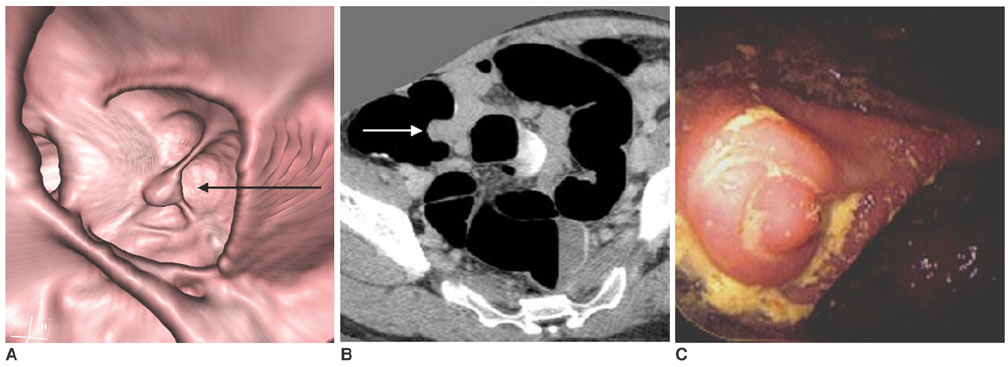
Fig. 2 A pseudopolyp at the appendiceal orifice in 35-year-old asymptomatic woman. A. A three-dimensional view of the cecal lumen revealing a 15 mm, polypoid lesion (arrow) at the appendiceal orifice. This lesion was misinterpreted as a cecal polyp on CT colonography. B. A contrast-enhanced axial CT image at the level of the cecum, shows an enhancing polypoid lesion (arrow) at the appendiceal orifice. This lesion was constant and non-movable in the prone, supine, and decubitus positions. C. A photograph from a conventional colonoscopy displaying the nodular endoluminal protrusion at the appendiceal base (not an intussusception of the appendix). The woman underwent pelvic surgery for a cesarean section and pelvic endometriosis a few years earlier. The endoluminal protrusion at the appendiceal base into the air-inflated cecum is thought to be due to pelvic adhesions.
Reference
-
1. Pickhardt PJ, Choi JR, Hwang I, Butler JA, Puckett ML, Hildebrandt HA, et al. Computed tomography virtual colonoscopy to screen for colorectal neoplasia in asymptomatic adults. N Engl J Med. 2003. 349:2191–2200.2. Johnson CD, Harmsen WS, Wilson LA, Maccarty RL, Welch TJ, Ilstrup DM, et al. Prospective blinded evaluation of computed tomographic colonography for screen detection of colorectal polyps. Gastroenterology. 2003. 125:311–319.3. Van Gelder RE, Nio CY, Florie J, Bartelsman JF, Snel P, De Jager SW, et al. Computed tomographic colonography compared with colonoscopy in patients at increased risk for colorectal cancer. Gastroenterology. 2004. 127:41–48.4. Cotton PB, Durkalski VL, Pineau BC, Palesch YY, Mauldin PD, Hoffman B, et al. Computed tomographic colonography: a multicenter comparison with standard colonoscopy for detection of colorectal neoplasia. JAMA. 2004. 291:1713–1719.5. Pineau BC, Paskett ED, Chen GJ, Espeland MA, Phillips K, Han JP, et al. Virtual colonoscopy using oral contrast compared with colonoscopy for the detection of patients with colorectal polyps. Gastroenterology. 2003. 125:304–310.6. Yee J, Akerkar GA, Hung RK, Steinauer-Gebauer AM, Wall SD, McQuaid KR. Colorectal neoplasia: performance characteristics of CT colonography for detection in 300 patients. Radiology. 2001. 219:685–692.7. Johnson CD, Toledano AY, Herman BA, Dachman AH, McFarland EG, Barish MA, et al. Computerized tomograhpic colonography: performance evaluation in a retrospective multicenter setting. Gastroenterology. 2003. 125:688–695.8. Spinzi G, Belloni G, Martegani A, Sangiovanni A, Favero CD, Minoli G. Computed tomographic colonography and conventional colonoscopy for colon diseases: a prospective, blinded study. Am J Gastroenterol. 2001. 96:394–400.9. Fenlon HM, Nunes DP, Schroy PC 3rd, Barish MA, Clarke PD, Ferrucci JT. A comparison of virtual and conventional colonoscopy for the detection of colorectal polyps. N Engl J Med. 1999. 341:1496–1503.10. Iannaccone R, Laghi A, Catalano C, Brink JA, Mangiapane F, Trenna S, et al. Detection of Colorectal lesions: lower-dose multi-detector row helical CT colonography compared with conventional colonoscopy. Radiology. 2003. 229:775–781.11. Macari M, Bini EJ, Xue X, Milano A, Katz SS, Resnick D, et al. Colorectal neoplasm: prospective comparison of thin-section low-dose multi-detector row CT colonography and conventional colonoscopy for detection. Radiology. 2002. 224:383–392.12. Durkalski VL, Palesch YY, Pineau BC, Vining DJ, Cotton PB. The virtual colonoscopy study: A large multicenter clinical trial designed to compare two diagnostic screening procedures. Control Clin Trials. 2002. 23:570–583.13. Kudo S, Kashida H, Tamura T, Koqure E, Imai Y, Yamano H, et al. Colonoscopic diagnosis and management of nonpolypoid early colorectal cancer. World J Surg. 2000. 24:1081–1090.14. Muto T, Kamiya J, Sawada T, Morioka Y. Morphogenesis of human colonic cancer. Dis Colon Rectum. 1983. 26:257–262.15. Lefere PA, Gryspeerdt SS, Dewyspelaere J, Baekelandt M, Van Holsbeeck BG. Dietary fecal tagging as a cleansing method before CT colonography: Initial results-polyp detection and patient acceptance. Radiology. 2002. 224:393–403.16. Lefere P, Gryspeerdt S, Marrannes J, Baekelandt M, Van Holsbeeck B. CT colonography after fecal tagging with a reduced cathartic cleansing and a reduced volume of barium. AJR Am J Roentgenol. 2005. 184:1836–1842.17. Iannaccone R, Laghi A, Catalano C, Mangiapane F, Lamazza A, Schillaci A, et al. Computed tomographic colonography without cathartic preparation for the detection of colorectal polyps. Gastroenterology. 2004. 127:1300–1311.18. Callstrom MR, Johnson CD, Fletcher JG, Reed JE, Ahlquist DA, Harmsen WS, et al. CT colonography without cathartic preparation: feasibility study. Radiology. 2001. 219:693–698.19. Lefere P, Gryspeerdt S, Baekelandt M, Van Holsbeeck B. Laxative-free CT colonography. AJR Am J Roentgenol. 2004. 183:945–948.20. Park SH, Ha HK, Kim MJ, Kim KW, Kim AY, Yang DH, et al. False-negative results at multi-defector row CT colonography: Multivariate analysis of causes for missed lesions. Radiology. 2005. 235:495–502.21. Postic G, Lewin D, Bickerstaff C, Wallace MB. Colonoscopic miss rates determined by direct comparison of colonoscopy with colon resection specimens. Am J Gastroenterol. 2002. 97:3182–3185.22. Rex DK, Cutler CS, Lemmel GT, Rahmani EY, Clark DW, Helper DJ, et al. Colonoscopic miss rates of adenomas determined by back-to-back colonoscopies. Gastroenterology. 1997. 112:24–28.23. Macari M, Bini EJ, Jacobs SL, Lui YW, Laks S, Milano A, et al. Significance of missed polyps at CT colonography. AJR Am J Roentgenol. 2004. 183:127–134.24. Gluecker TM, Fletcher JG, Welch TJ, MacCarty RL, Harmsen WS, Harrington JR, et al. Characterization of lesions missed on interpretation of CT colonography using a 2D search method. AJR Am J Roentgenol. 2004. 182:881–889.25. Rembacken BJ, Fujii T, Cairns A, Dixon MF, Yoshida S, Chalmers DM, et al. Flat and depressed colonic neoplasms: a prospective study of 1000 colonoscopies in the UK. Lancet. 2000. 355:1211–1214.26. Hurlstone DP, Cross SS, Adam I, Shorthouse AJ, Brown S, Sanders DS, et al. A prospective clinicopathological and endoscopic evaluation of flat and depressed colorectal lesions in the United Kingdom. Am J Gastroenterol. 2003. 98:2543–2549.
- Full Text Links
-
- Actions
-
Cited
- CITED
-
- Close
- Share
-
- Similar articles
-
- Efficacy of CT Colonography in the Detection of Colorectal Polypoid Lesions
- Utility of CT colonography in detecting colon polyps as a colon cancer screen
- CT Colonography
- Can Computed Tomography Colonography Replace Optical Colonoscopy in Detecting Colorectal Lesions?: State of the Art
- Virtual CT Colonoscopy and Virtual CT Barium Enema using Multidetector-row CT
