Efficacy of Barium-Based Fecal Tagging for CT Colonography: a Comparison between the Use of High and Low Density Barium Suspensions in a Korean Population - a Preliminary Study
- Affiliations
-
- 1Department of Radiology and Research Institute of Radiology and Internal Medicine, University of Ulsan College of Medicine, Asan Medical Center, Seoul, Korea. seongho@amc.seoul.kr
- 2Department of Radiology, Asan Medical Center, Seoul, Korea.
- 3Department of Radiology, David Geffen School of Medicine, University of California Los Angeles, Los Angeles, CA, USA.
- 4National Cancer Center, Gyeonggi-do, Korea.
- KMID: 1088675
- DOI: http://doi.org/10.3348/kjr.2009.10.1.25
Abstract
OBJECTIVE
This preliminarily study was designed to determine and to compare the efficacy of two commercially available barium-based fecal tagging agents for CT colonography (CTC) (high-density [40% w/v] and low-density [4.6% w/v] barium suspensions) in a population in Korea. MATERIALS AND METHODS: In a population with an identified with an average-risk for colorectal cancer, 15 adults were administered three doses of 20 ml 40% w/v barium for fecal tagging (group I) and 15 adults were administered three doses of 200 ml 4.6% w/v barium (group II) for fecal tagging. Excluding five patients in group I and one patient in group II that left the study, ten patients in group I and 14 patients in group II were finally included in the analysis. Two experienced readers evaluated the CTC images in consensus regarding the degree of tagging of stool pieces 6 mm or larger. Stool pieces were confirmed with the use of standardized CTC criteria or the absence of matched lesions as seen on colonoscopy. The rates of complete fecal tagging were analyzed on a per-lesion and a per-segment basis and were compared between the patients in the two groups. RESULTS: Per-lesion rates of complete fecal tagging were 52% (22 of 42; 95% CI, 37.7-66.6%) in group I and 78% (28 of 36; 95% CI, 61.7-88.5%) in group II. The difference between the two groups did not reach statistical significance (p = 0.285). The per-segment rates of complete tagging were 33% (6 of 18; 95% CI, 16.1%-56.4%) in group I and 60% (9 of 15; 95% CI, 35.7%-80.3%) in group II; again, the difference between the two groups did not reach statistical significance (p = 0.171). CONCLUSION: Barium-based fecal tagging using both the 40% w/v and the 4.6% w/v barium suspensions showed moderate tagging efficacy. The preliminary comparison did not demonstrate a statistically significant difference in the tagging efficacy between the use of the two tagging agents, despite the tendency toward better tagging with the use of the 4.6% w/v barium suspension.
Keyword
MeSH Terms
Figure
-
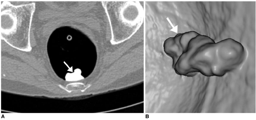
Fig. 1 Presence of completely tagged stool piece in 62-year-old man. A. Transverse 2D CT colonography image of colon window setting (width, 1,500H; level, -200H) obtained with supine position shows 44-mm completely tagged stool piece of homogeneous high attenuation (arrow) in rectum. B. 3D endoluminal CT colonography image shows same stool piece (arrow) as in A.
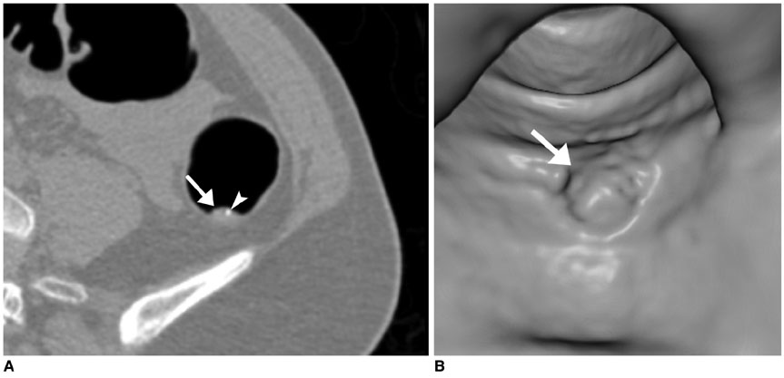
Fig. 2 Incompletely tagged stool piece in 55-year-old woman. A. Transverse 2D CT colonography image of colon window setting (width, 1,500H; level, -200H) obtained with patient in supine position shows 12-mm incompletely tagged stool piece (arrow) in descending colon. Only small fraction of stool piece is tagged (arrowhead). B. 3D endoluminal CT colonography image shows same stool piece (arrow) as in A.
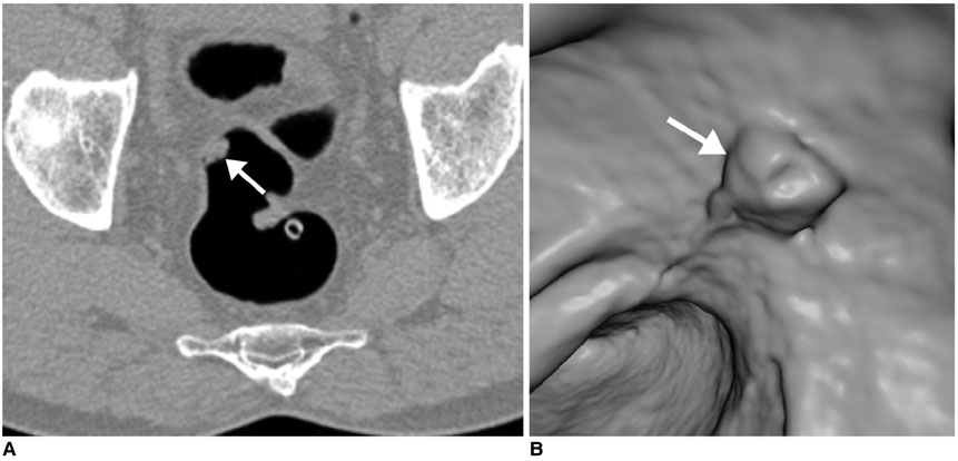
Fig. 3 Untagged stool piece in 58-year-old man. A. Transverse 2D CT colonography image of colon window setting (width, 1,500H; level, -200H) obtained with patient in prone position shows 14-mm untagged stool piece (arrow) in upper rectum, which shows similar attenuation to that of soft-tissue. B. 3D endoluminal CT colonography image shows same stool piece (arrow) as in A.
Reference
-
1. Park SH, Yee J, Kim SH, Kim YH. Fundamental elements for successful performance of CT colonography (virtual colonoscopy). Korean J Radiol. 2007. 8:264–275.2. Lefere PA, Gryspeerdt SS, Dewyspelaere J, Baekelandt M, Van Holsbeeck BG. Dietary fecal tagging as a cleansing method before CT colonography: initial results polyp detection and patient acceptance. Radiology. 2002. 224:393–403.3. Beebe TJ, Johnson CD, Stoner SM, Anderson KJ, Limburg PJ. Assessing attitudes toward laxative preparation in colorectal cancer screening and effects on future testing: potential receptivity to computed tomographic colonography. Mayo Clin Proc. 2007. 82:666–671.4. Ristvedt SL, McFarland EG, Weinstock LB, Thyssen EP. Patient preferences for CT colonography, conventional colonoscopy, and bowel preparation. Am J Gastroenterol. 2003. 98:578–585.5. Bielen D, Thomeer M, Vanbeckevoort D, Kiss G, Maes F, Marchal G, et al. Dry preparation for virtual CT colonography with fecal tagging using water-soluble contrast medium: initial results. Eur Radiol. 2003. 13:453–458.6. Callstrom MR, Johnson CD, Fletcher JG, Reed JE, Ahlquist DA, Harmsen WS, et al. CT colonography without cathartic preparation: feasibility study. Radiology. 2001. 219:693–698.7. Dachman AH, Dawson DO, Lefere P, Yoshida H, Khan NU, Cipriani N, et al. Comparison of routine and unprepped CT colonography augmented by low fiber diet and stool tagging: A pilot study. Abdom Imaging. 2007. 32:96–104.8. Gryspeerdt S, Lefere P, Herman M, Deman R, Rutgeerts L, Ghillebert G, et al. CT colonography with fecal tagging after incomplete colonoscopy. Eur Radiol. 2005. 15:1192–1202.9. Iannaccone R, Laghi A, Catalano C, Mangiapane F, Lamazza A, Schillaci A, et al. Computed tomographic colonography without cathartic preparation for the detection of colorectal polyps. Gastroenterology. 2004. 127:1300–1311.10. Johnson CD, Manduca A, Fletcher JG, MacCarty RL, Carston MJ, Harmsen WS, et al. Noncathartic CT colonography with stool tagging: performance with and without electronic stool subtraction. AJR Am J Roentgenol. 2008. 190:361–366.11. Johnson KT, Carston MJ, Wentz RJ, Manduca A, Anderson SM, Johnson CD. Development of a cathartic-free colorectal cancer screening test using virtual colonoscopy: a feasibility study. AJR Am J Roentgenol. 2007. 188:W29–W36.12. Lefere P, Gryspeerdt S, Baekelandt M, Van Holsbeeck B. Laxative-free CT colonography. AJR Am J Roentgenol. 2004. 183:945–948.13. Lefere P, Gryspeerdt S, Marrannes J, Baekelandt M, Van Holsbeeck B. CT colonography after fecal tagging with a reduced cathartic cleansing and a reduced volume of barium. AJR Am J Roentgenol. 2005. 184:1836–1842.14. Pickhardt PJ, Choi JR, Hwang I, Butler JA, Puckett ML, Hildebrandt HA, et al. Computed tomographic virtual colonoscopy to screen for colorectal neoplasia in asymptomatic adults. N Engl J Med. 2003. 349:2191–2200.15. An S, Lee KH, Kim YH, Park SH, Kim HY, Kim SH, et al. Screening CT colonography in an asymptomatic average-risk Asian population: a 2-year experience in a single institution. AJR Am J Roentgenol. 2008. 191:W100–W106.16. Yun JY, Ro HJ, Park JB, Choi JB, Chung JE, Kim YJ, et al. Diagnostic performance of CT colonography for the detection of colorectal polyps. Korean J Radiol. 2007. 8:484–491.17. Dictionary.Com. Accessed February 21, 2008. http://dictionary.reference.com/browse/Kimchi.htm.18. Dachman AH, Zalis ME. Quality and consistency in CT colonography and research reporting. Radiology. 2004. 230:319–323.19. Taylor SA, Halligan S, Goh V, Morley S, Bassett P, Atkin W, et al. Optimizing colonic distention for multi-detector row CT colonography: effect of hyoscine butylbromide and rectal balloon catheter. Radiology. 2003. 229:99–108.20. Pickhardt PJ, Lee AD, McFarland EG, Taylor AJ. Linear polyp measurement at CT colonography: in vitro and in vivo comparison of two-dimensional and three-dimensional displays. Radiology. 2005. 236:872–878.21. Lee SS, Park SH, Choi EK, Kim SY, Kim MJ, Lee KH, et al. Colorectal polyps on portal phase contrast-enhanced CT colonography: lesion attenuation and distinction from tagged feces. AJR Am J Roentgenol. 2007. 189:35–40.22. Taylor SA, Slater A, Burling DN, Tam E, Greenhalgh R, Gartner L, et al. CT colonography: optimisation, diagnostic performance and patient acceptability of reduced-laxative regimens using barium-based faecal tagging. Eur Radiol. 2008. 18:32–42.23. Best preps are tailored to VC reading method. AuntMinnie.com. 2005. 11. 16. Accessed August 6, 2007. http://www.auntminnie.com.24. Park SH, Lee SS, Kim JK, Kim MJ, Kim HJ, Kim SY, et al. Volume rendering with color coding of tagged stool during endoluminal fly-through CT colonography: effect on reading efficiency. Radiology. 2008. 248:1018–1027.25. Pickhardt PJ. Screening CT colonography: how I do it. AJR Am J Roentgenol. 2007. 189:290–298.26. Yoshida H, Nappi J. CAD in CT colonography without and with oral contrast agents: progress and challenges. Comput Med Imaging Graph. 2007. 31:267–284.27. New Zealand medicines and medical devices safety authority. Medsafe website. Accessed August 10, 2007. http://www.medsafe.govt.nz.
- Full Text Links
-
- Actions
-
Cited
- CITED
-
- Close
- Share
-
- Similar articles
-
- CT Colonography
- Virtual CT Colonoscopy and Virtual CT Barium Enema using Multidetector-row CT
- Acute Respiratory Failure Caused by Aspiration of High Density Barium: A Case Report
- Comparison of pressure between barium reduction and air reduction of the intussusception in children
- Strategy for early detection of colon cancer
