Photodynamic Therapy Treatment for Eyes with Drusenoid Pigment Epithelium Detachment
- Affiliations
-
- 1Department of Ophthalmology, College of Medicine, The Catholic University of Korea, Seoul, Korea.
- 2Department of Ophthalmology, Saevit Eye Hospital, Gyeonggi-do, Korea. kiseok-kim@daum.net
- KMID: 1070758
- DOI: http://doi.org/10.3341/kjo.2008.22.3.194
Abstract
- We report the clinical course of photodynamic therapy (PDT) in a patient with drusenoid pigment epithelium detachment (PED). A patient with drusenoid PED underwent PDT follow-up was carried out at one week, one month, three months, six months and one year after treatment. Fundus exam, optical coherence tomography (OCT) and fluorescein angiography were performed. After the PDT, drusen and PED were gradually diminished over one year. However, pure serous PED eventually developed at the same location of the drusenoid PED. The results of the PDT, on drusenoid PED, were initially effective, but not completely successful. Therefore, PDT may be considered as an alternative treatment option for drusenoid PED.
MeSH Terms
Figure
-
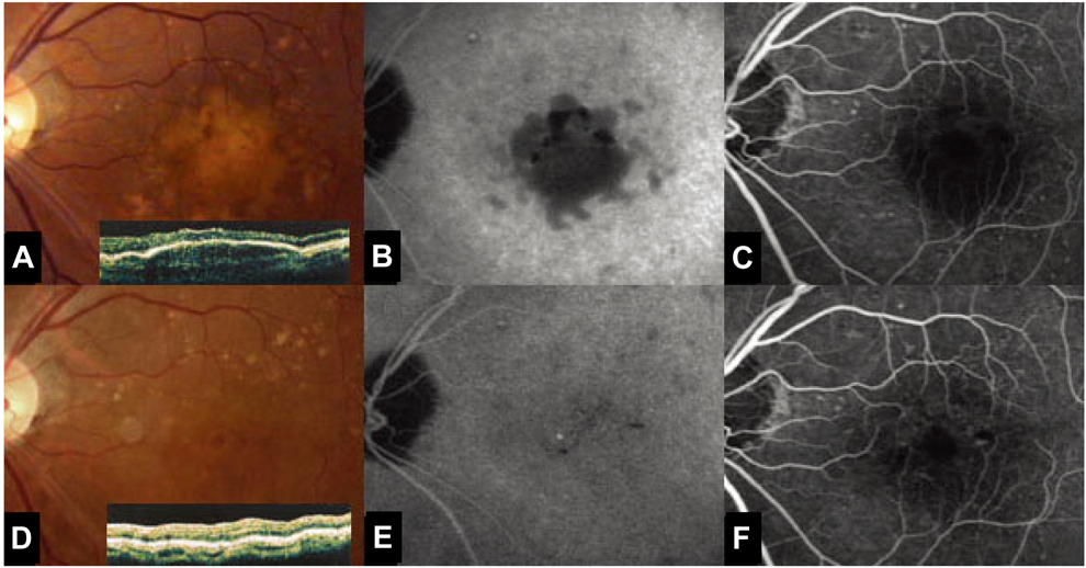
Fig. 1 (A, B, C) Fundus photography and optical coherence tomography (OCT) at the first examination showed multiple pigment epithelium detachments. Fluorescein angiography revealed delayed, regular hyperfluorescence without leakage. There was no evidence of choroidal neovascularization (CNV). (D, E, F) Ten months after photodynamic therapy (PDT), fundus photography, OCT and ICG showed that the drusenoid PED diminished in size.
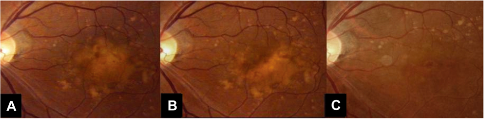
Fig. 2 (A) Fifteen days after PDT, there was no interval change, on the fundus examination. (B) Five months after PDT, the fundus examination showed a decreased size of the soft drusen. (C) Ten months after PDT, the corrected vision of the left eye was improved to 20/25. On fundus examination, the number of soft drusen was markedly decreased.
Reference
-
1. Ryan SJ. Retina. 2006. Vol. 2:4th ed. Philadelphia: Elsevier mosby;1083.2. Photodynamic therapy of subfovealchoroidal neovascularization in age-related macular degeneration with verteporfin: one-year results of 2 randomized clinical trials-TAP report. Treatment of age-related macular degeneration with photodynamic therapy (TAP) Study Group. Arch Ophthalmol. 1999. 117:1329–1345.3. Roquet W, Roudot-Thoraval F, Coscas G, Soubrane G. Clinical features of drusenoid pigment epithelial detachment in age related macular degeneration. Br J Ophthalmol. 2004. 88:638–642.4. Ho AC, Maguire MG, Yoken J, et al. Laser-induced drusen reduction improves visual function at 1 year. Ophthalmology. 1999. 106:1367–1374.5. Verteporfin In Photodynamic Therapy Study Group. Verteporfin therapy of subfoveal choroidal neovascularization in age-related macular degeneration: two-year results of a randomized clinical trial including lesions with occult with no classic choroidal neovascularization-verteporfin in photodynamic therapy report 2. Am J Ophthalmol. 2001. 131:541–560.6. Frennesson C, Nilsson SE. Prophylactic laser treatment in early age related maculopathy reduced the incidence of exudative complications. Br J Ophthalmol. 1998. 82:1169–1174.7. Pauleikhoff D, Löffert D, Spital G, et al. Pigment epithelial detachment in the elderly. Clinical differentiation, natural course and pathogenetic implications. Graefe's Arch Clin Exp Ophthalmol. 2002. 240:533–538.
- Full Text Links
-
- Actions
-
Cited
- CITED
-
- Close
- Share
-
- Similar articles
-
- Photodynamic Therapy of Choroidal Neovasculariation Associated with Large Serous Pigment Epithelial Detachment
- Photodynamic Therapy with Verteporfin in Polypoidal Choroidal Vasculopathy
- Morphological Changes of Retinal Pigment Epithelium After Experimental Retinal Detachment
- Combination Treatment for Choroidal Neovascularization Associated With Large Retinal Pigment Epithelial Detachment
- The Effect of Photodynamic Therapy in Chronic Central Serous Chorioretinopathy
