Korean J Ophthalmol.
2011 Dec;25(6):380-386. 10.3341/kjo.2011.25.6.380.
Photoreceptor Disruption Related to Persistent Submacular Fluid after Successful Scleral Buckle Surgery
- Affiliations
-
- 1Department of Ophthalmology, Seoul National University Bundang Hospital, Seoul National University College of Medicine, Seongnam, Korea. jiani4@snu.ac.kr
- 2Department of Ophthalmology, Seoul National University Hospital, Seoul National University College of Medicine, Seoul, Korea.
- KMID: 1031184
- DOI: http://doi.org/10.3341/kjo.2011.25.6.380
Abstract
- PURPOSE
To investigate serial changes in photoreceptor status and associated visual outcome in patients with persistent submacular fluid after successful scleral buckle surgery for macula-off rhegmatogenous retinal detachment.
METHODS
This was a prospective observational case series including 76 consecutive patients who underwent successful scleral buckle surgery for macula-off rhegmatogenous retinal detachment with symptom duration < or =90 days at a single tertiary hospital. Optical coherence tomography (OCT) and visual acuity examination were performed at one month and three months postoperatively and at three-month intervals until the submacular fluid disappeared. Main outcome measures were postoperative photoreceptor status on OCT and visual acuity.
RESULTS
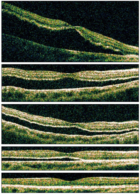
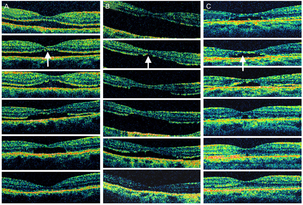
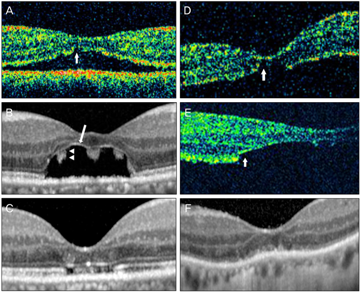
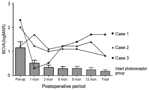
Forty-two patients (55.3%) showed persistent submacular fluid at postoperative one month. Of 42 patients with persistent submacular fluid, three (7.1%) showed photoreceptor disruption on OCT. None of the 34 patients without persistent submacular fluid showed photoreceptor disruption. Two patients (4.8%) had progressive photoreceptor disruption, and one patient (2.4%) had early photoreceptor disruption. All three patients showed photoreceptor reappearance and limited visual restoration after absorption of submacular fluid. Final visual acuities were significantly worse in these three patients (20 / 1000, 20 / 133, and 20 / 133) compared to those of the other patients (mean, 20 / 30) with persistent submacular fluid and intact photoreceptors.
CONCLUSIONS
Even after successful scleral buckle surgery for rhegmatogenous retinal detachment, photoreceptor disruption can occur related to persistent submacular fluid and may be a cause of poor visual outcome.
Keyword
MeSH Terms
Figure
Reference
-
1. Wolfensberger TJ, Gonvers M. Optical coherence tomography in the evaluation of incomplete visual acuity recovery after macula-off retinal detachments. Graefes Arch Clin Exp Ophthalmol. 2002. 240:85–89.2. Seo JH, Woo SJ, Park KH, et al. Influence of persistent submacular fluid on visual outcome after successful scleral buckle surgery for macula-off retinal detachment. Am J Ophthalmol. 2008. 145:915–922.3. Hagimura N, Iida T, Suto K, Kishi S. Persistent foveal retinal detachment after successful rhegmatogenous retinal detachment surgery. Am J Ophthalmol. 2002. 133:516–520.4. Benson SE, Schlottmann PG, Bunce C, et al. Optical coherence tomography analysis of the macula after scleral buckle surgery for retinal detachment. Ophthalmology. 2007. 114:108–112.5. Ross WH. Visual recovery after macula-off retinal detachment. Eye (Lond). 2002. 16:440–446.6. Smith AJ, Telander DG, Zawadzki RJ, et al. High-resolution Fourier-domain optical coherence tomography and microperimetric findings after macula-off retinal detachment repair. Ophthalmology. 2008. 115:1923–1929.7. Wakabayashi T, Oshima Y, Fujimoto H, et al. Foveal microstructure and visual acuity after retinal detachment repair: imaging analysis by Fourier-domain optical coherence tomography. Ophthalmology. 2009. 116:519–528.8. Suh MH, Seo JM, Park KH, Yu HG. Associations between macular findings by optical coherence tomography and visual outcomes after epiretinal membrane removal. Am J Ophthalmol. 2009. 147:473–480.e3.9. Matsumoto H, Kishi S, Otani T, Sato T. Elongation of photoreceptor outer segment in central serous chorioretinopathy. Am J Ophthalmol. 2008. 145:162–168.10. Maruko I, Iida T, Sekiryu T, Saito M. Morphologic changes in the outer layer of the detached retina in rhegmatogenous retinal detachment and central serous chorioretinopathy. Am J Ophthalmol. 2009. 147:489–494.e1.11. Berglin L, Algvere PV, Seregard S. Photoreceptor decay over time and apoptosis in experimental retinal detachment. Graefes Arch Clin Exp Ophthalmol. 1997. 235:306–312.12. Mervin K, Valter K, Maslim J, et al. Limiting photoreceptor death and deconstruction during experimental retinal detachment: the value of oxygen supplementation. Am J Ophthalmol. 1999. 128:155–164.13. Lee SY, Joe SG, Kim JG, et al. Optical coherence tomography evaluation of detached macula from rhegmatogenous retinal detachment and central serous chorioretinopathy. Am J Ophthalmol. 2008. 145:1071–1076.14. Immonen I, Konttinen YT, Sorsa T, et al. Proteinases in subretinal fluid. Graefes Arch Clin Exp Ophthalmol. 1996. 234:105–109.15. Symeonidis C, Diza E, Papakonstantinou E, et al. Expression of matrix metalloproteinases in the subretinal fluid correlates with the extent of rhegmatogenous retinal detachment. Graefes Arch Clin Exp Ophthalmol. 2007. 245:560–568.16. Hewitt AT, Lindsey JD, Carbott D, Adler R. Photoreceptor survival-promoting activity in interphotoreceptor matrix preparations: characterization and partial purification. Exp Eye Res. 1990. 50:79–88.17. Yang L, Bula D, Arroyo JG, Chen DF. Preventing retinal detachment-associated photoreceptor cell loss in Bax-deficient mice. Invest Ophthalmol Vis Sci. 2004. 45:648–654.18. Curcio CA, Sloan KR, Kalina RE, Hendrickson AE. Human photoreceptor topography. J Comp Neurol. 1990. 292:497–523.
- Full Text Links
- Actions
-
Cited
- CITED
-
- Close
- Share
- Similar articles
-
- Recurrence of Retinal Detachment after Scleral Buckle Removal
- Effect of Transscleral Diode Laser Photocoagulation Applied Through Silicone Scleral Exoplants
- Geometric changes of the eye with an encircling scleral buckle
- Clinical Evaluation of Ptosis after Scleral Buckling
- Drainge vs. Nondrainage of Suvretinal Fluid in Scleral Buckling Procedure


