Clinical Application of the Unifying Concept of Cutaneous Melanoma
- Affiliations
-
- 1Provincial Health Care Services, Institute of Pathology, Santa Maria del Carmine Hospital, Rovereto (TN), Italy. emailmedical@gmail.com
- 2Department of Diagnostic and Clinical Medicine and of Public Health, Section of Pathology, University of Modena and Reggio Emilia, Modena (MO), Italy.
- KMID: 2367344
- DOI: http://doi.org/10.4068/cmj.2017.53.1.78
Abstract
- No abstract available.
MeSH Terms
Figure
-
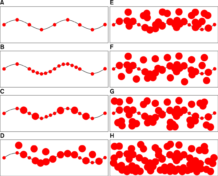
FIG. 1 Schematic representation of the progression from normal skin to invasive malignant melanoma. (A) Normal skin with melanocytes (smaller grey bullets) regularly distributed at the basis of the epidermis (black line). (B) Epidermis with basal hyperplastic melanocytes. (C) Epidermis with basal atypical melanocytes (larger grey bullets). (D) Intra-epidermal radial growth phase of primary malignant melanoma with lentiginous pattern. (E) Intra-epidermal radial growth phase of primary malignant melanoma with pagetoid pattern. (F) Micro-invasive radial growth phase of primary malignant melanoma with lentiginous pattern (single melanoma cells are present in the papillary dermis). (G) Micro-invasive radial growth phase of primary malignant melanoma with pagetoid pattern (small clusters of melanoma cells are present in the papillary dermis). (H) Invasive (early if ≤1 mm in thick) vertical growth phase of primary cutaneous malignant melanoma (large clusters of melanoma cells are present in the papillary dermis and beyond).
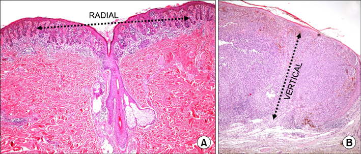
FIG. 2 Histopathological exemplification of the radial growth phase (A: hematoxylin/eosin, 4×) and of the vertical one (B: hematoxylin/eosin, 4×) in primary cutaneous malignant melanoma.
Cited by 1 articles
-
The ‘All-or-None Law’ Applied to the Vertical Growth Phase of Cutaneous Malignant Melanoma
Luca Roncati, Benedetto Vergari, Antonio Del Gaudio
Chonnam Med J. 2017;53(3):234-235. doi: 10.4068/cmj.2017.53.3.234.
Reference
-
1. Kim SY, Yun SJ. Cutaneous melanoma in Asians. Chonnam Med J. 2016; 52:185–193.
Article2. Ackerman AB. Malignant melanoma: a unifying concept. Hum Pathol. 1980; 11:591–595.
Article3. Roncati L, Piscioli F, Pusiol T. Sentinel lymph node in thin and thick melanoma. Klin Onkol. 2016; 29:393–394.4. Piscioli F, Pusiol T, Roncati L. Wisely choosing thin melanomas for sentinel lymph node biopsy. J Am Acad Dermatol. 2017; 76:e25.
Article5. Madu MF, Wouters MW, van Akkooi AC. Sentinel node biopsy in melanoma: current controversies addressed. Eur J Surg Oncol. 2016; DOI: 10.1016/j.ejso.2016.08.007. [Epub ahead of print].
Article6. Piscioli F, Pusiol T, Roncati L. Histopathological determination of thin melanomas at risk for metastasis. Melanoma Res. 2016; 26:635.
Article7. Piscioli F, Pusiol T, Roncati L. Nowadays a histological sub-typing of thin melanoma is demanded for a proper patient management. J Plast Reconstr Aesthet Surg. 2016; 69:1563–1564.
Article8. Pusiol T, Piscioli F, Speziali L, Zorzi MG, Morichetti D, Roncati L. Clinical features, dermoscopic patterns, and histological diagnostic model for melanocytic tumors of uncertain malignant potential (MELTUMP). Acta Dermatovenerol Croat. 2015; 23:185–194.9. Elder DE, Xu X. The approach to the patient with a difficult melanocytic lesion. Pathology. 2004; 36:428–434.
Article10. Piscioli F, Pusiol T, Roncati L. Diagnostic approach to melanocytic lesion of unknown malignant potential. Melanoma Res. 2016; 26:91–92.
Article11. Piscioli F, Pusiol T, Roncati L. Diagnostic disputes regarding atypical melanocytic lesions can be solved by using the term MELTUMP. Turk Patoloji Derg. 2016; 32:63–64.
Article12. Piscioli F, Pusiol T, Roncati L. Thin melanoma subtyping fits well with the American Joint Committee on Cancer staging system. Melanoma Res. 2016; 26:636.
Article13. Roncati L, Piscioli F, Pusiol T. Surgical outcomes reflect the histological types of cutaneous malignant melanoma. J Eur Acad Dermatol Venereol. 2016; DOI: 10.1111/jdv.14023. [Epub ahead of print].
Article
- Full Text Links
-
- Actions
-
Cited
- CITED
-
- Close
- Share
-
- Similar articles
-
- Cutaneous Malignant Melanoma Associated with Papillary Thyroid Cancer
- A Case of Cutaneous Metastatic Melanoma Treated with Topical Diphencyprone (DPCP)
- Cutaneous Melanoma in Asians
- Cutaneous Ulceration after Injection of Interferon Alpha in a Melanoma Patient
- Primary cutaneous malignant melanoma of the breast
