Expression of ciliary neurotrophic factor and its receptor in experimental obstructive nephropathy
- Affiliations
-
- 1Department of Anatomy, College of Medicine, The Catholic University of Korea, Seoul, Korea. jhcha@catholic.ac.kr
- KMID: 1447418
- DOI: http://doi.org/10.5115/acb.2011.44.2.85
Abstract
- Ciliary neurotrophic factor (CNTF) is well known as a growth/survival factor of neuronal tissue. We investigated the expression of CNTF and its specific receptor alpha (CNTFRalpha) in a unilateral ureteral obstruction (UUO) model. Complete UUO was produced by left ureteral ligation in Sprague-Dawley rats. The animals were sacrificed on days 1, 3, 5, 7, 14, 21, and 28 after UUO. The kidneys were fixed, and processed for both immunohistochemistry and in situ hybridization. CNTF immunoreactivity in sham-operated kidneys was observed only in the descending thin limb (DTL) of the loop of Henle. In UUO kidneys, CNTF expression was induced in the S3 segment (S3s) of the proximal tubule from day 1, and progressively expanded into the entire S3s and a part of the convoluted proximal tubules, distal tubules (DT), and glomerular parietal epithelium up to day 7. Upregulated CNTF expression was maintained to day 28. From day 14, the inner medullary collecting duct showed weak CNTF immunoreactivity. The CNTFRalpha mRNA hybridization signal in sham-operated kidneys was weakly detected in the DTL, DT, medullary thick ascending limb, and in a few S3s cells. After UUO, CNTFRalpha mRNA expression increased progressively in both the renal cortex and the medulla up to day 7 and increased expression was maintained until day 28. The results suggest that the S3s may be the principal induction site for CNTF in response to renal injury, and that CNTF may play a role in chronic renal injury.
Keyword
MeSH Terms
-
Animals
Chimera
Ciliary Neurotrophic Factor
Ciliary Neurotrophic Factor Receptor alpha Subunit
Epithelium
Extremities
Immunohistochemistry
In Situ Hybridization
Kidney
Ligation
Loop of Henle
Neurons
Rats, Sprague-Dawley
RNA, Messenger
Ureter
Ureteral Obstruction
Ciliary Neurotrophic Factor
Ciliary Neurotrophic Factor Receptor alpha Subunit
RNA, Messenger
Figure
-
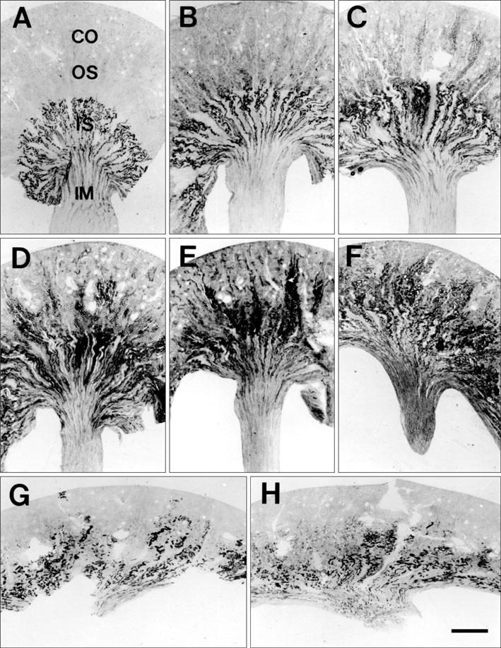
Fig. 1 Light micrographs of a 50-µm thick vibratome section illustrating ciliary neurotrophic factor (CNTF) immunostaining in kidney of sham-operated rat (A), and in kidneys of rats who underwent unilateral ureteral obstruction (UUO) on days 1 (B), 3 (C), 5 (D), 7 (E), 14 (F), 21 (G), and 28 (H). (A) In sham-operated rats, CNTF-immunostaining was strong in the inner stripe (IS) of the outer medulla and in the inner medulla (IM). (B) From day 1 after UUO, induced CNTF immunostaining started to appear between the outer stripe (OS) of the outer medulla and the cortex (CO). CNTF immunostaining expanded into the cortex and the outer stripe of the outer medulla by day 7 (C-E), and then decreased slightly (F-H), but CNTF immunostaining in the OS and IS remained strong up to day 28 after UUO. Scale bar=1,000 µm.
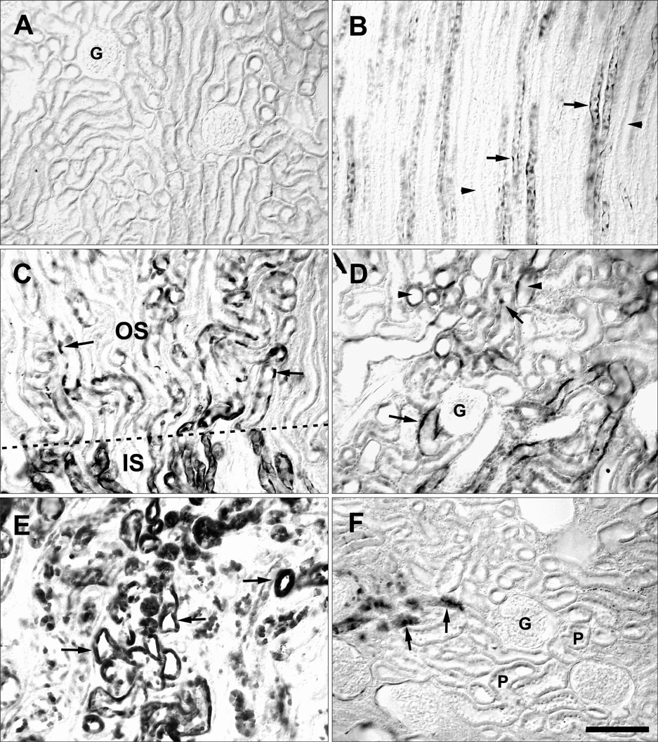
Fig. 2 High magnification of sham-operated (A, B) kidneys and those of rats who underwent unilateral ureteral obstruction on days 1 (C), 5 (D), 14 (E), and 28 (F) illustrating ciliary neurotrophic factor (CNTF) immunostaining. (A) No positive reaction was detected in the cortex. (B) Only the descending thin limb (arrows) showed CNTF immunoreactivity in the inner medulla . The collecting ducts (arrowheads) were negative. CNTF immunoreactivity was induced in several cells (arrows in C) of the S3 segment. Dotted lines indicate the boundary between the outer stripe (OS) and inner stripe (IS). Many CNTF-positive distal tubules (arrowheads) and proximal tubular cells (arrows) could be seen on day 5 (D), but only small numbers of tubular profiles (arrows) were immunoreactive on day 28 (F) in the cortex. (E) Note the numerous S3 segments with intense immunoreactivity (arrows). G, glomerulus; P, proximal tubule. Scale bar=100 µm.
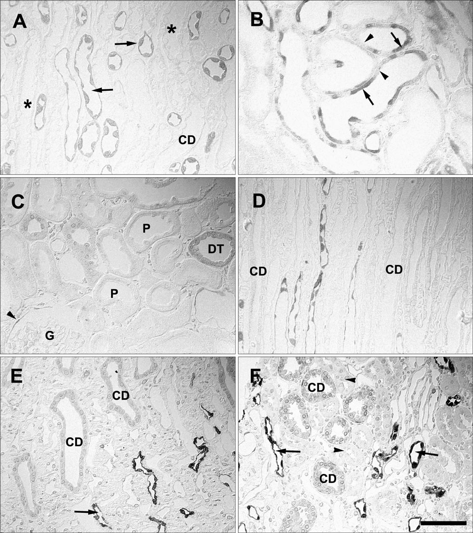
Fig. 3 One-µm thick sections from sham-operated (A) kidney and those of rats who underwent unilateral ureteral obstruction on days 7 (B), 21 (C, F), 3 (D), and 14 (E) illustrating ciliary neurotrophic factor (CNTF) immunostaining. (A) The descending thin limb (arrows) showed CNTF immunoreactivity in the outer medulla, whereas the thick ascending limb (asterisks) and collecting duct (CD) were negative. Note the CNTF-positive cells with no brush border (arrows in B) and CNTF-negative cells with a well-developed brush border (arrowheads in B) in the S3 segments. Cortical immunoreactivity on day 21 (C) was usually detected in the distal tubule (DT) and parietal epithelium (arrowhead) of Bowman's capsule. Proximal tubules (Ps in C) were immunonegative and appeared morphologically normal. The inner medullary collecting duct (CDs in D-F) showed no immunoreactivity up to day 7 (D), but weak immunostaining was detected from day 14 (E, F). Note strong immunoreactivity in the descending thin limb (arrows in E and F) and no immunoreactivity in the thick ascending limb (arrowheads in F). G, glomerulus. Scale bar=50 µm.
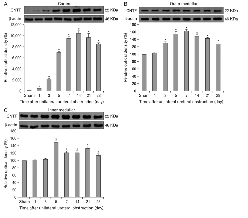
Fig. 4 Immunoblotting for ciliary neurotrophic factor (CNTF) protein in kidneys of rats after a sham operation or unilateral ureteral obstruction. A kidney from each rat was dissected into cortex, outer medulla, inner medulla, and protein (20 µg) was applied to each line. A 22-kDa band corresponded to the molecular weight of the CNTF protein. β-actin was re-probed to demonstrate equal protein loading and the 46-kDa band corresponded to the molecular weight. (A) Immunoblot for the CNTF protein in the cortex. (B) Immunoblot for the CNTF protein in the outer medulla. (C) Immunoblot for the CNTF protein in the inner medulla. *P<0.05 vs. sham. Optical densities are mean±standard deviation.
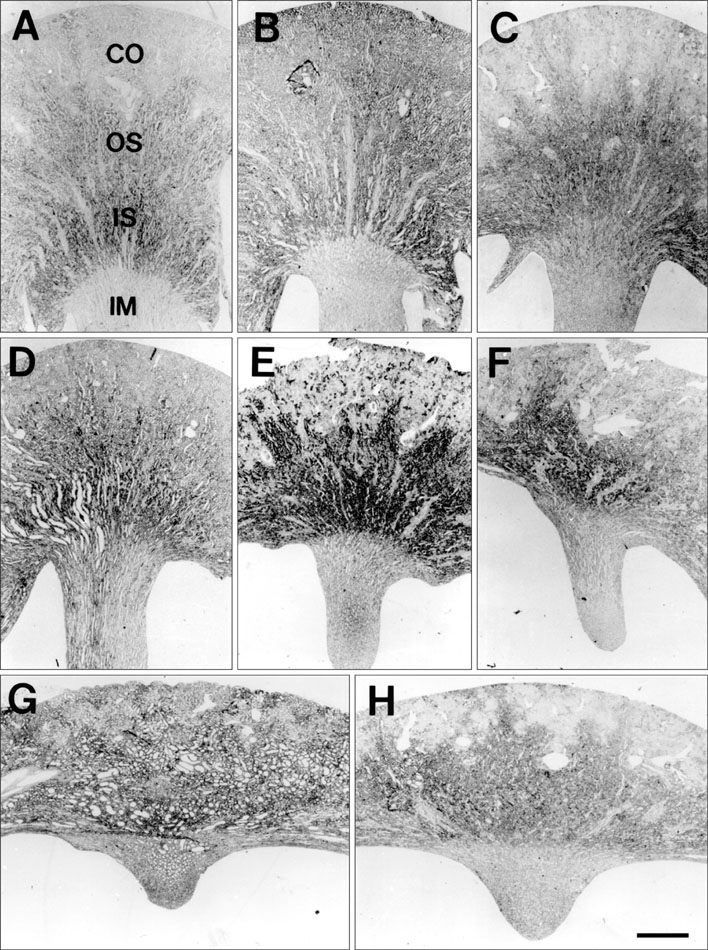
Fig. 5 Five µm thick waxed section illustrating ciliary neurotrophic factor-specific receptor alpha (CNTFRα) mRNA expression in kidney from sham-operated rat (A), and kidneys of rats who underwent unilateral ureteral obstruction on days 1 (B), 3 (C), 5 (D), 7 (E), 14 (F), 21 (G), and 28 (H). (A) A weak hybridization signal was observed in the cortex and outer medulla of sham-operated rats. In situ CNTFRα mRNA signals were induced in the outer medulla on day 1 (B), and the signals increased and expanded into the cortex on day 7 (C-E). Thereafter, the in situ CNTFRα mRNA signals decreased slightly (F-H). Note the persistently high levels of CNTFRα mRNA on day 28 (H). CO, cortex; OS, outer stripe; IS, inner stripe; IM, inner medulla. Scale bar=1,000 µm.
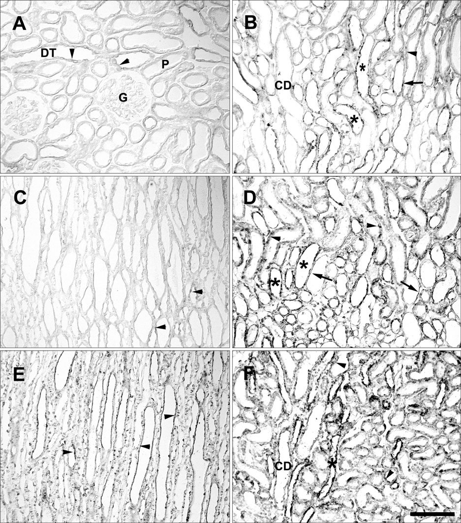
Fig. 6 High magnification of sham-operated (A-C) kidneys and those who underwent unilateral ureteral obstruction (D-F) illustrating ciliary neurotrophic factor-specific receptor alpha mRNA expression. A weak signal was shown only in some cells of the cortex (arrowheads in A) and of the distal tubule (DT). (B) Note the moderate to weak in situ signal in the thick ascending limb (asterisks), collecting duct (CD) in the outer stripe of outer medulla, and in the changing section from the descending thin limb to the S3 segment. The descending thin limb (arrow) was positive, whereas the S3 segment (arrowhead) was negative. (C) No detectable signal was observed in the inner medulla or the descending thin limb (arrowheads). On days 1 (D) and 3 (F), in the outer stripe of the outer medulla, moderately increased signals were observed in the thick ascending limb (asterisks), descending thin limb (arrows), and S3 segments (arrowheads). (E) The CDs of the inner medulla showed a moderate signal on day 1. G, glomerulus; P, proximal tubule. Scale bar=100 µm.
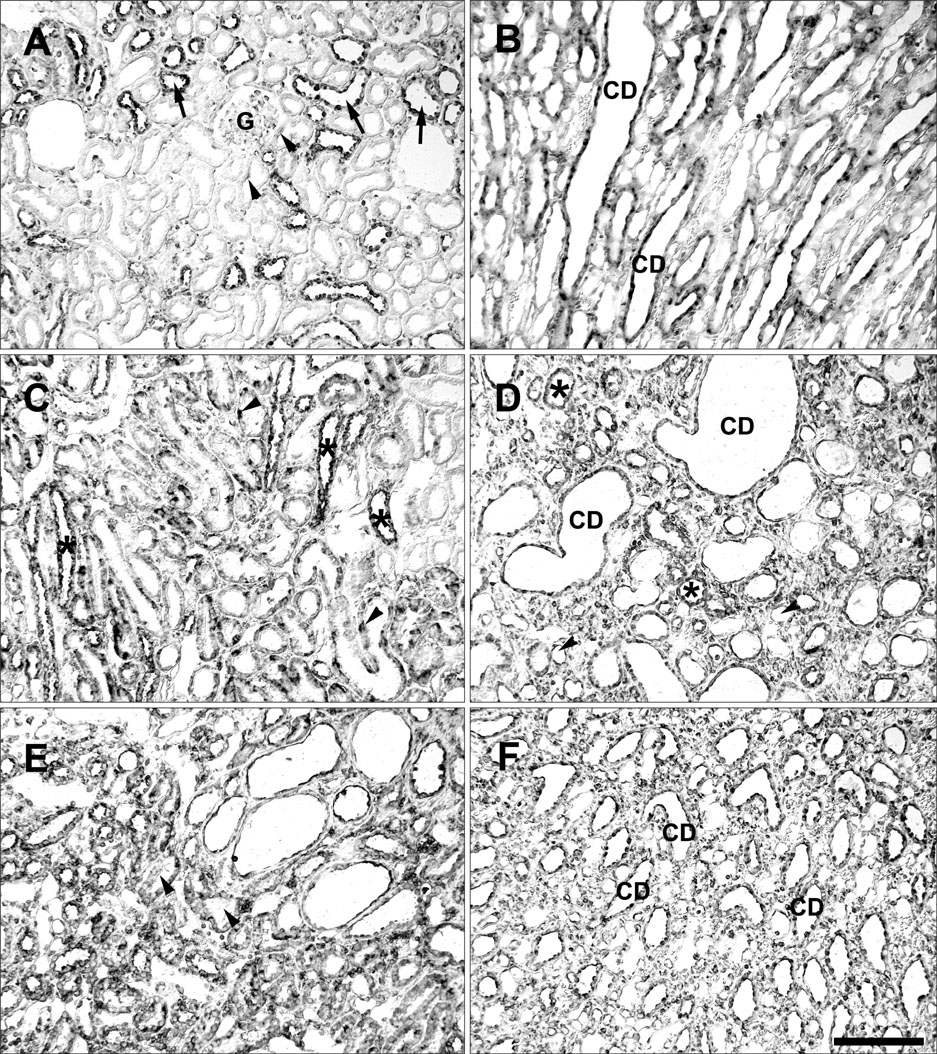
Fig. 7 High magnification of a rat kidney following unilateral ureteral obstruction illustrating ciliary neurotrophic factor-specific receptor alpha mRNA expression. (A) In the cortex, a strong signal was observed in the distal tubule (arrows), some proximal tubules (arrowheads), and in glomerular parietal epithelial cells (G) on day 5. In the outer stripe of the outer medulla on days 14 (C) and 28 (E), strong in situ signals were observed in the thick ascending limbs (asterisks) and S3 segments (arrowheads). (D) In the inner stripe of the outer medulla on day 21, signals were observed in the descending thin limb (arrowheads), thick ascending limb (asterisks), and collecting duct (CD). In the inner medulla, the CD showed a moderate signal on days 7 (B) and 28 (F). Scale bar=100 µm.
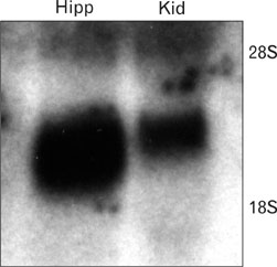
Fig. 8 Northern blot using a P32-labeled antisense ciliary neurotrophic factor-specific receptor alpha riboprobe. Total RNA (each 20 µg) from hippocampus (hipp) and renal medulla (kid) were separated.
Reference
-
1. Adler R, Landa KB, Manthorpe M, Varon S. Cholinergic neuronotrophic factors: intraocular distribution of trophic activity for ciliary neurons. Science. 1979. 204:1434–1436.2. Ip NY, Nye SH, Boulton TG, Davis S, Taga T, Li Y, Birren SJ, Yasukawa K, Kishimoto T, Anderson DJ, Stahl N, Yancopoulos GD. CNTF and LIF act on neuronal cells via shared signaling pathways that involve the IL-6 signal transducing receptor component gp130. Cell. 1992. 69:1121–1132.3. Ikeda K, Iwasaki Y, Shiojima T, Kinoshita M. Neuroprotective effect of various cytokines on developing spinal motoneurons following axotomy. J Neurol Sci. 1996. 135:109–113.4. Humes HD, Lake EW, Liu S. Renal tubule cell repair following acute renal injury. Miner Electrolyte Metab. 1995. 21:353–365.5. Heinrich PC, Behrmann I, Müller-Newen G, Schaper F, Graeve L. Interleukin-6-type cytokine signalling through the gp130/Jak/STAT pathway. Biochem J. 1998. 334(Pt 2):297–314.6. Davis S, Aldrich TH, Stahl N, Pan L, Taga T, Kishimoto T, Ip NY, Yancopoulos GD. LIFR beta and gp130 as heterodimerizing signal transducers of the tripartite CNTF receptor. Science. 1993. 260:1805–1808.7. Ohta K, Hara H, Hayashi K, Itoh N, Ohi T, Ohta M. Tissue expression of rat ciliary neurotrophic factor (CNTF) mRNA and production of the recombinant CNTF. Biochem Mol Biol Int. 1995. 35:283–290.8. Ohta M, Ohi T, Nishimura M, Itoh N, Hayashi K, Ohta K. Distribution of and age-related changes in ciliary neurotrophic factor protein in rat tissues. Biochem Mol Biol Int. 1996. 40:671–678.9. Ong AC, Fine LG. Tubular-derived growth factors and cytokines in the pathogenesis of tubulointerstitial fibrosis: implications for human renal disease progression. Am J Kidney Dis. 1994. 23:205–209.10. Yang CW, Lim SW, Han KW, Ahn HJ, Park JH, Kim YH, Kirsh M, Cha JH, Park JH, Kim YS, Kim J, Bang BK. Upregulation of ciliary neurotrophic factor (CNTF) and CNTF receptor alpha in rat kidney with ischemia-reperfusion injury. J Am Soc Nephrol. 2001. 12:749–757.11. Kim J, Kim YH, Cha JH, Tisher CC, Madsen KM. Intercalated cell subtypes in connecting tubule and cortical collecting duct of rat and mouse. J Am Soc Nephrol. 1999. 10:1–12.12. Davis S, Aldrich TH, Valenzuela DM, Wong VV, Furth ME, Squinto SP, Yancopoulos GD. The receptor of ciliary neurotrophic factor. Science. 1991. 253:59–63.13. Truong LD, Petrusevska G, Yang G, Gurpinar T, Shappell S, Lechago J, Rouse D, Suki WN. Cell apoptosis and proliferation in experimental chronic obstructive uropathy. Kidney Int. 1996. 50:200–207.14. Nguyen HT, Wu HY, Baskin LS, Kogan BA. High urinary flow accelerates renal injury in young rats with partial unilateral ureteral obstruction. J Urol. 2000. 163:1904–1907.15. Adler R. Ciliary neurotrophic factor as an injury factor. Curr Opin Neurobiol. 1993. 3:785–789.16. Sleeman MW, Anderson KD, Lambert PD, Yancopoulos GD, Wiegand SJ. The ciliary neurotrophic factor and its receptor, CNTFR alpha. Pharm Acta Helv. 2000. 74:265–272.17. Stöckli KA, Lottspeich F, Sendtner M, Masiakowski P, Carroll P, Götz R, Lindholm D, Thoenen H. Molecular cloning, expression and regional distribution of rat ciliary neurotrophic factor. Nature. 1989. 342:920–923.18. Lillien LE, Sendtner M, Rohrer H, Hughes SM, Raff MC. Type-2 astrocyte development in rat brain cultures is initiated by a CNTF-like protein produced by type-1 astrocytes. Neuron. 1988. 1:485–494.19. Lin LF, Mismer D, Lile JD, Armes LG, Butler ET 3rd, Vannice JL, Collins F. Purification, cloning, and expression of ciliary neurotrophic factor (CNTF). Science. 1989. 246:1023–1025.20. Kamiguchi H, Yoshida K, Sagoh M, Sasaki H, Inaba M, Wakamoto H, Otani M, Toya S. Release of ciliary neurotrophic factor from cultured astrocytes and its modulation by cytokines. Neurochem Res. 1995. 20:1187–1193.21. Lee MY, Naumann T, Kirsch M, Frotscher M, Hofmann HD. Transient up-regulation of ciliary neurotrophic factor receptor-alpha mRNA in axotomized rat septal neurons. Eur J Neurosci. 1997. 9:622–626.22. Chevalier RL, Goyal S, Wolstenholme JT, Thornhill BA. Obstructive nephropathy in the neonatal rat is attenuated by epidermal growth factor. Kidney Int. 1998. 54:38–47.23. Chevalier RL, Goyal S, Kim A, Chang AY, Landau D, LeRoith D. Renal tubulointerstitial injury from ureteral obstruction in the neonatal rat is attenuated by IGF-1. Kidney Int. 2000. 57:882–890.24. Truong LD, Gaber L, Eknoyan G. Obstructive uropathy. Contrib Nephrol. 2011. 169:311–326.25. Elbjeirami WM, Truong LD, Tawil A, Wang W, Dawson S, Lan HY, Zhang P, Garcia GE, Wayne Smith C. Early differential expression of oncostatin M in obstructive nephropathy. J Interferon Cytokine Res. 2010. 30:513–523.26. Akin M, Demirbilek S, Ay S, Gurunluoglu K, Turkmen E, Tas E, Aksoy RT, Baykarabulut A, Edali MN. Attenuation of ureteral obstruction-induced renal injury by polyenyl phos phatidylcholine. Int J Urol. 2007. 14:350–356.
- Full Text Links
-
- Actions
-
Cited
- CITED
-
- Close
- Share
-
- Similar articles
-
- Polymorphism of the Ciliary Neurotrophic Factor (CNTF) Gene in Korean
- microRNA-146a Promotes Growth of Acute Leukemia Cells by Downregulating Ciliary Neurotrophic Factor Receptor and Activating JAK2/STAT3 Signaling
- Delayed Treatment of Capsaicin Produces Partial Motor Recovery by Enhancing Dopamine Function in MPPâº-lesioned Rats via Ciliary Neurotrophic Factor
- The Effect of Recombinant Tyrosine Hydroxylase Expression on the Neurogenic Differentiation Potency of Mesenchymal Stem Cells
- Neuronal Rescue by Neurotrophic Factors in Human Fetal Cerebral Neuron Cultures Exposed to Oxygen Radical Injury
