Neuroprotective Effects of Sacral Epidural Neuromodulation Following Spinal Cord Injury : An Experimental Study in Rats
- Affiliations
-
- 1Department of Neurosurgery, Seoul National University Bundang Hospital, Seoul National University College of Medicine, Seongnam, Korea. neurospine@snubh.org
- 2Department of Urology, Seoul National University Bundang Hospital, Seoul National University College of Medicine, Seongnam, Korea.
- 3Department of Rehabilitation Medicine, Seoul National University Bundang Hospital, Seoul National University College of Medicine, Seongnam, Korea.
- KMID: 1426258
- DOI: http://doi.org/10.3340/jkns.2012.52.6.509
Abstract
OBJECTIVE
The purpose of this study is to evaluate neuroprotective effect of sacral neuromodulation in rat spinal cord injury (SCI) model in the histological and functional aspects.
METHODS
Twenty-one female Sprague Dawley rats were randomly divided into 3 groups : the normal control group (CTL, n=7), the SCI with sham stimulation group (SCI, n=7), and the SCI with electrical stimulation (SCI+ES, n=7). Spinal cord was injured by dropping an impactor from 25 mm height. Sacral nerve electrical stimulation was performed by the following protocol : pulse duration, 0.1 ms; frequency, 20 Hz; stimulation time, 30 minutes; and stimulation duration, 4 weeks. Both locomotor function and histological examination were evaluated as scheduled.
RESULTS
The number of anterior horn cell was 12.3+/-5.7 cells/high power field (HPF) in the CTL group, 7.8+/-4.9 cells/HPF in the SCI group, and 6.9+/-5.5 cells/HPF in the SCI+ES group, respectively. Both the SCI and the SCI+ES groups showed severe loss of anterior horn cells and myelin fibers compared with the CTL group. Cavitation and demyelinization of the nerve fibers has no significant difference between the SCI group and the SCI+ES group. Cavitation of dorsal column was more evident in only two rats of SCI group than the SCI+ES group. The locomotor function of all rats improved over time but there was no significant difference at any point in time between the SCI and the SCI+ES group.
CONCLUSION
In a rat thoracic spinal cord contusion model, we observed that sacral neuromodulation did not prevent SCI-induced myelin loss and apoptosis.
MeSH Terms
Figure
-
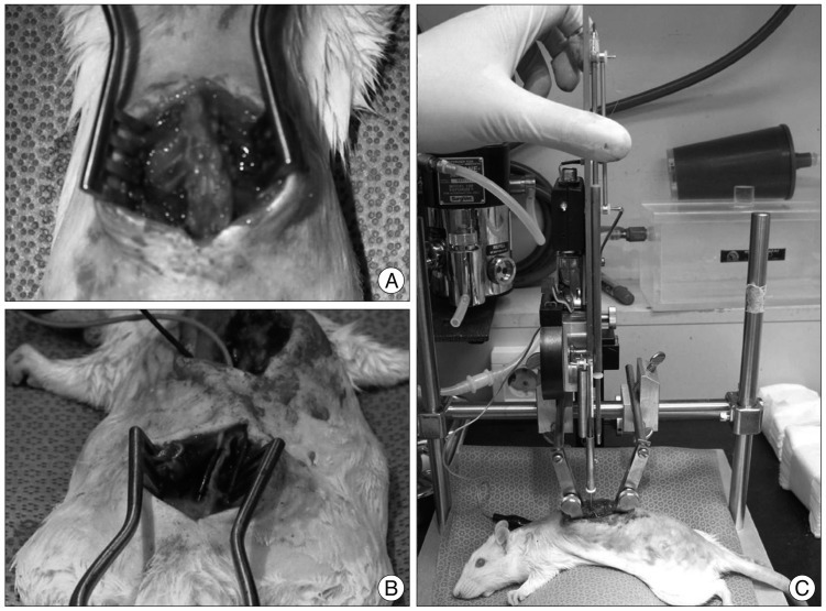
Fig. 1 A : Exposure of spinal cord at T10-11 by laminectomy. B : Fine needle electrodes (0.5×27 G) are implanted into S2 foramen. C : Severe grade of crushing injury is made using New York University spinal cord impactor.

Fig. 2 A : Hematoxylin and eosin staining of control group. Dorsal column is intact. There are many ant horn cells (arrows) in gray matter. B : The spinal cord injury (SCI) group reveals a cavitation of dorsal column and a loss of anterior horn cell (arrows) were observed. C : The SCI+ES group shows no structural defect, but loss of anterior horn cell (arrows) is similar with the SCI group.

Fig. 3 A : Luxol fast blue staining of control group. Myelin fibers are stained. B : The spinal cord injury (SCI) group shows structural defect and scant myelin fibers. C : The SCI+ES group also reveals cavitation of dorsal column with vacuolar change and loss of myelin fibers. Structural defect and cavitation of dorsal column is more evident in SCI group.

Fig. 4 The time course of Basso, Beattie, and Bresnahan (BBB) scores in the spinal cord injury (SCI) and SCI+ES group. All rats show a low BBB score at 1 day after SCI then recovered slowly. The difference between the groups is not significant.
Reference
-
1. Agrawal G, Kerr C, Thakor NV, All AH. Characterization of graded multicenter animal spinal cord injury study contusion spinal cord injury using somatosensory-evoked potentials. Spine (Phila Pa 1976). 2010; 35:1122–1127. PMID: 20354478.
Article2. Al-Majed AA, Neumann CM, Brushart TM, Gordon T. Brief electrical stimulation promotes the speed and accuracy of motor axonal regeneration. J Neurosci. 2000; 20:2602–2608. PMID: 10729340.
Article3. Bai C, An H, Wang S, Jiang D, Fan W, Nie H. Treatment and prevention of bacterial translocation and endotoxemia with stimulation of the sacral nerve root in a rabbit model of spinal cord injury. Spine (Phila Pa 1976). 2011; 36:363–371. PMID: 20531069.
Article4. Basso DM, Beattie MS, Bresnahan JC. A sensitive and reliable locomotor rating scale for open field testing in rats. J Neurotrauma. 1995; 12:1–21. PMID: 7783230.
Article5. Basso DM, Beattie MS, Bresnahan JC. Graded histological and locomotor outcomes after spinal cord contusion using the NYU weight-drop device versus transection. Exp Neurol. 1996; 139:244–256. PMID: 8654527.
Article6. Brindley GS, Polkey CE, Rushton DN, Cardozo L. Sacral anterior root stimulators for bladder control in paraplegia : the first 50 cases. J Neurol Neurosurg Psychiatry. 1986; 49:1104–1114. PMID: 3491180.
Article7. Cohen DM, Patel CB, Ahobila-Vajjula P, Sundberg LM, Chacko T, Liu SJ, et al. Blood-spinal cord barrier permeability in experimental spinal cord injury : dynamic contrast-enhanced MRI. NMR Biomed. 2009; 22:332–341. PMID: 19023867.
Article8. Ebner A, Jiang C, Lindström S. Intravesical electrical stimulation--an experimental analysis of the mechanism of action. J Urol. 1992; 148:920–924. PMID: 1512860.
Article9. Geremia NM, Gordon T, Brushart TM, Al-Majed AA, Verge VM. Electrical stimulation promotes sensory neuron regeneration and growth-associated gene expression. Exp Neurol. 2007; 205:347–359. PMID: 17428474.
Article10. Hausmann ON. Post-traumatic inflammation following spinal cord injury. Spinal Cord. 2003; 41:369–378. PMID: 12815368.
Article11. Hong CH, Lee HY, Jin MH, Noh JY, Lee BH, Han SW. The effect of intravesical electrical stimulation on bladder function and synaptic neurotransmission in the rat spinal cord after spinal cord injury. BJU Int. 2009; 103:1136–1141. PMID: 19021629.
Article12. Inoue M, Hojo T, Yano T, Katsumi Y. The effects of electroacupuncture on peripheral nerve regeneration in rats. Acupunct Med. 2003; 21:9–17. PMID: 12924841.
Article13. Ishigooka M, Suzuki Y, Hashimoto T, Sasagawa I, Nakada T, Handa Y. A new technique for sacral nerve stimulation : a percutaneous method for urinary incontinence caused by spinal cord injury. Br J Urol. 1998; 81:315–318. PMID: 9488079.
Article14. Kim KT, Nam TK, Park YS, Kim YB, Park SW. Neuroprotective effect of anthocyanin on experimental traumatic spinal cord injury. J Korean Neurosurg Soc. 2011; 49:205–211. PMID: 21607177.
Article15. Lee TT, Green BA, Dietrich WD, Yezierski RP. Neuroprotective effects of basic fibroblast growth factor following spinal cord contusion injury in the rat. J Neurotrauma. 1999; 16:347–356. PMID: 10369555.
Article16. O'Gara T, Urban W, Polishchuk D, Pierre-Louis A, Stewart M. Continuous stimulation of transected distal nerves fails to prolong action potential propagation. Clin Orthop Relat Res. 2006; 447:209–213. PMID: 16505717.17. Rijkhoff NJ, Wijkstra H, van Kerrebroeck PE, Debruyne FM. Selective detrusor activation by electrical sacral nerve root stimulation in spinal cord injury. J Urol. 1997; 157:1504–1508. PMID: 9120991.
Article
- Full Text Links
-
- Actions
-
Cited
- CITED
-
- Close
- Share
-
- Similar articles
-
- Comparative Analysis Between Thoracic Spinal Cord and Sacral Neuromodulation in a Rat Spinal Cord Injury Model: A Preliminary Report of a Rat Spinal Cord Stimulation Model
- The effects of nalbuphine and fentanyl on experimental spinal cord injuries
- Effects of Methylprednisolone on Neuroprotective Effects of Delay Hypothermia on Spinal Cord Injury in Rat
- Failure of topical DMSO to improve blood flow or evoked potentials in rat spinal cord injury
- The Effect of the Swimming Exercise on Functional Recovery after Experimental Contusive Spinal Cord Injury in the Rats
