Maturation of bone marrow-derived dendritic cells by a novel beta-glucan purified from Paenibacillus polymyxa JB115
- Affiliations
-
- 1College of Veterinary Medicine, Jeju National University, Jeju 690-756, Korea. jooh@jejunu.ac.kr
- 2College of Veterinary Medicine, Kyungpook National University, Daegu 702-701, Korea.
- 3College of Veterinary Medicine, Kangwon National University, Chuncheon 200-701, Korea.
- KMID: 1106562
- DOI: http://doi.org/10.4142/jvs.2011.12.2.187
Abstract
- We investigated the immunostimulatory effects of a novel beta-glucan purified from Paenibacillus (P.) polymyxa JB115 on bone marrow-derived dendritic cells (DCs), a type of potent antigen-presenting cells. beta-glucan isolated from P. polymyxa JB115 enhanced the viability and induced the maturation of DCs. beta-glucan markedly increased the cytokine production of DCs and surface expression of DC markers. In addition, DCs treated with beta-glucan showed a higher capacity to stimulate allogeneic spleen cell proliferation compared to those treated with medium alone. These results demonstrate the effect of beta-glucan on DC maturation and may increase the use of beta-glucan.
Keyword
MeSH Terms
-
Animals
Bone Marrow Cells/cytology/*drug effects/*immunology
Cell Survival/drug effects/*immunology
Dendritic Cells/cytology/*drug effects/*immunology
Flow Cytometry
Immunophenotyping/methods
Interleukin-12/analysis/immunology
Mice
Mice, Inbred BALB C
Nitric Oxide/analysis/immunology
Paenibacillus/*chemistry
Tumor Necrosis Factor-alpha/analysis/immunology
beta-Glucans/isolation & purification/*pharmacology
Figure
-
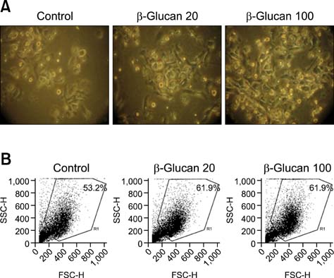
Fig. 1 Clusters of dendritic cells (DCs) treated with β-glucan. DCs were seeded at a density of 5 × 10⁵ cells/mL in 6-well culture plates, and then treated with β-glucan for 2 days. (A) DC morphology. ×200. (B) The number and size of DCs was measured by flow cytometric analysis.

Fig. 2 Effect of β-glucan on DC viability. DCs were seeded at a concentration of 2.5 × 10⁵ cells/mL in 96-well culture plates, and then treated with the indicated concentrations of β-glucan for 2 days. For the viability assay, treated cells were stained with a trypan blue staining solution to determine the number of live and dead cells. Data are presented as the mean ± SD from four individual wells. **p < 0.01.
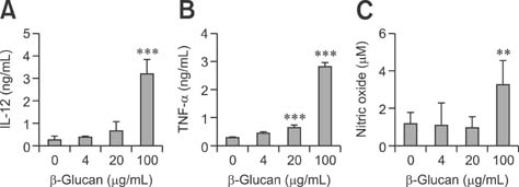
Fig. 3 β-glucan increases cytokine and nitric oxide production of DCs. DCs were seeded and treated as described in Fig. 2. The amounts of interleukin (IL)-12 (A), tumor necrosis factor (TNF)-α (B), and nitric oxide (C) produced by DCs were measured. Data are presented as the mean ± SD from four individual wells. **p < 0.01, ***p < 0.001.
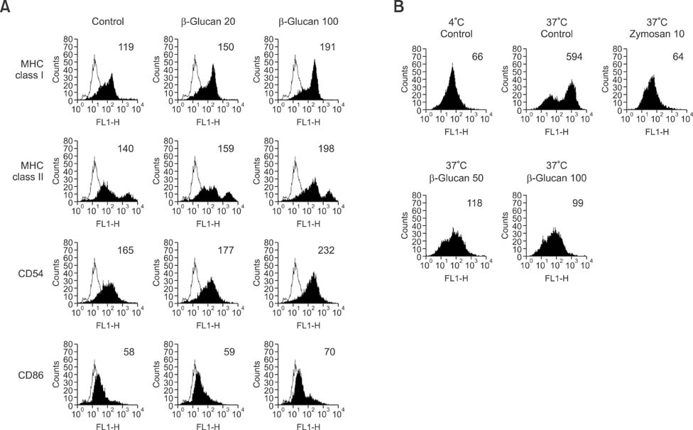
Fig. 4 β-glucan enhances the expression of immune-related DC surface markers but decreases DC antigen uptake. DCs were prepared and treated as described in the legend for Fig. 1. For surface marker analysis (A), the number in the histogram indicates the mean fluorescence intensity (MFI) of the main DC population. For antigen uptake analysis (B), DCs were incubated with 250 µg/mL dextran-fluorescein isothiocyanate at 4℃ or 37℃ for 45 min. The number in the histogram indicates the MFI of viable DCs.
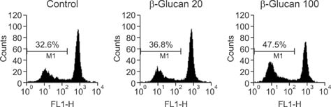
Fig. 5 β-glucan increases the allo-proliferative capability of DCs. DC cultures were established and treated as described in the legend for Fig. 1. For the allo-proliferation assay, DCs treated with β-glucan (1 × 10⁴ cells/well) were co-cultured for 5 days with allogeneic splenocytes (2 × 10⁵ cells/well) stained with 5 µM carboxyfluorescein succinimidyl ester. The number indicates the percentage of proliferating cells with a low FL1 value in the co-culture.
Cited by 1 articles
-
Distinct Effects of Monophosphoryl Lipid A, Oligodeoxynucleotide CpG, and Combination Adjuvants on Modulating Innate and Adaptive Immune Responses to Influenza Vaccination
Eun-Ju Ko, Young-Tae Lee, Youri Lee, Ki-Hye Kim, Sang-Moo Kang
Immune Netw. 2017;17(5):326-342. doi: 10.4110/in.2017.17.5.326.
Reference
-
1. Brown GD, Gordon S. Immune recognition of fungal β-glucans. Cell Microbiol. 2005. 7:471–479.
Article2. Chang ZQ, Lee JS, Gebru E, Hong JH, Jung HK, Jo WS, Park SC. Mechanism of macrophage activation induced by β-glucan produced from Paenibacillus polymyxa JB115. Biochem Biophys Res Commun. 2010. 391:1358–1362.
Article3. Esche C, Shurin GV, Kirkwood JM, Wang GQ, Rabinowich H, Pirtskhalaishvili G, Shurin MR. Tumor necrosis factor-α-promoted expression of Bcl-2 and inhibition of mitochondrial cytochrome c release mediate resistance of mature dendritic cells to melanoma-induced apoptosis. Clin Cancer Res. 2001. 7:3 Suppl. 974s–979s.4. Goodridge HS, Wolf AJ, Underhill DM. β-glucan recognition by the innate immune system. Immunol Rev. 2009. 230:38–50.
Article5. Heufler C, Koch F, Stanzl U, Topar G, Wysocka M, Trinchieri G, Enk A, Steinman RM, Romani N, Schuler G. Interleukin-12 is produced by dendritic cells and mediates T helper 1 development as well as interferon-γ production by T helper 1 cells. Eur J Immunol. 1996. 26:659–668.
Article6. Inaba K, Witmer-Pack M, Inaba M, Hathcock KS, Sakuta H, Azuma M, Yagita H, Okumura K, Linsley PS, Ikehara S, Muramatsu S, Hodes RJ, Steinman RM. The tissue distribution of the B7-2 costimulator in mice: abundant expression on dendritic cells in situ and during maturation in vitro. J Exp Med. 1994. 180:1849–1860.
Article7. Joo HG, Goedegebuure PS, Sadanaga N, Nagoshi M, von Bernstorff W, Eberlein TJ. Expression and function of galectin-3, a β-galactoside-binding protein in activated T lymphocytes. J Leukoc Biol. 2001. 69:555–564.8. Jung HK, Hong JH, Park SC, Park BK, Nam DH, Kim SD. Production and physicochemical characterization of β-glucan produced by Paenibacillus polymyxa JB115. Biotechnol Bioprocess Eng. 2007. 12:713–719.
Article9. Kim MH, Byon YY, Ko EJ, Song JY, Yun YS, Shin T, Joo HG. Immunomodulatory activity of ginsan, a polysaccharide of Panax ginseng, on dendritic cells. Korean J Physiol Pharmacol. 2009. 13:169–173.
Article10. Kim MH, Joo HG. Immunostimulatory effects of fucoidan on bone marrow-derived dendritic cells. Immunol Lett. 2008. 115:138–143.
Article11. Larsen CP, Ritchie SC, Hendrix R, Linsley PS, Hathcock KS, Hodes RJ, Lowry RP, Pearson TC. Regulation of immunostimulatory function and costimulatory molecule (B7-1 and B7-2) expression on murine dendritic cells. J Immunol. 1994. 152:5208–5219.12. Mellman I, Steinman RM. Dendritic cells: specialized and regulated antigen processing machines. Cell. 2001. 106:255–258.13. Steinman RM, Pack M, Inaba K. Dendritic cells in the T-cell areas of lymphoid organs. Immunol Rev. 1997. 156:25–37.
Article
- Full Text Links
-
- Actions
-
Cited
- CITED
-
- Close
- Share
-
- Similar articles
-
- Immunostimulatory Effects of beta-glucan Purified from Paenibacillus polymyxa JB115 on Mouse Splenocytes
- A novel beta-glucan produced by Paenibacillus polymyxa JB115 induces nitric oxide production in RAW264.7 macrophages
- Defects in the differentiation and function of bone marrow-derived dendritic cells in non-obese diabetic mice
- Immunomodulatory Effects of Eckol, a Pure Compound of Ecklonia Cava, on Dendritic Cells
- Mycobacterium tuberculosis ESAT6 Drives the Activation and Maturation of Bone Marrow-Derived Dendritic Cells via TLR4-Mediated Signaling
