MR Imaging Findings of a Primary Cardiac Osteosarcoma and Its Bone Metastasis with Histopathologic Correlation
- Affiliations
-
- 1Department of Radiology and Institute of Radiation Medicine, Seoul National University College of Medicine, Seoul 110-744, Korea. jacrad@radiol.snu.ac.kr
- 2Department of Radiology, Seoul National University Bundang Hospital, Gyeonggi-do 463-707, Korea.
- 3Department of Pathology, Seoul National University Bundang Hospital, Gyeonggi-do 463-707, Korea.
- 4Department of Radiology, Gyeongsang National University Hospital, Jinju 660-702, Korea.
- KMID: 991692
- DOI: http://doi.org/10.3348/kjr.2011.12.1.135
Abstract
- An osteosarcoma of cardiac origin is extremely rare, and a comprehensive description of MR imaging (MRI) findings of cardiac osteosarcoma and its metastasis in the femur have not been reported in the literature. We present a case of cardiac osteosarcoma in a 47-year-old woman and its metastasis to the femur, focusing on the description of MRI findings of the cardiac and metastatic bony osteosarcoma with a histopathologic correlation.
Keyword
MeSH Terms
Figure
-
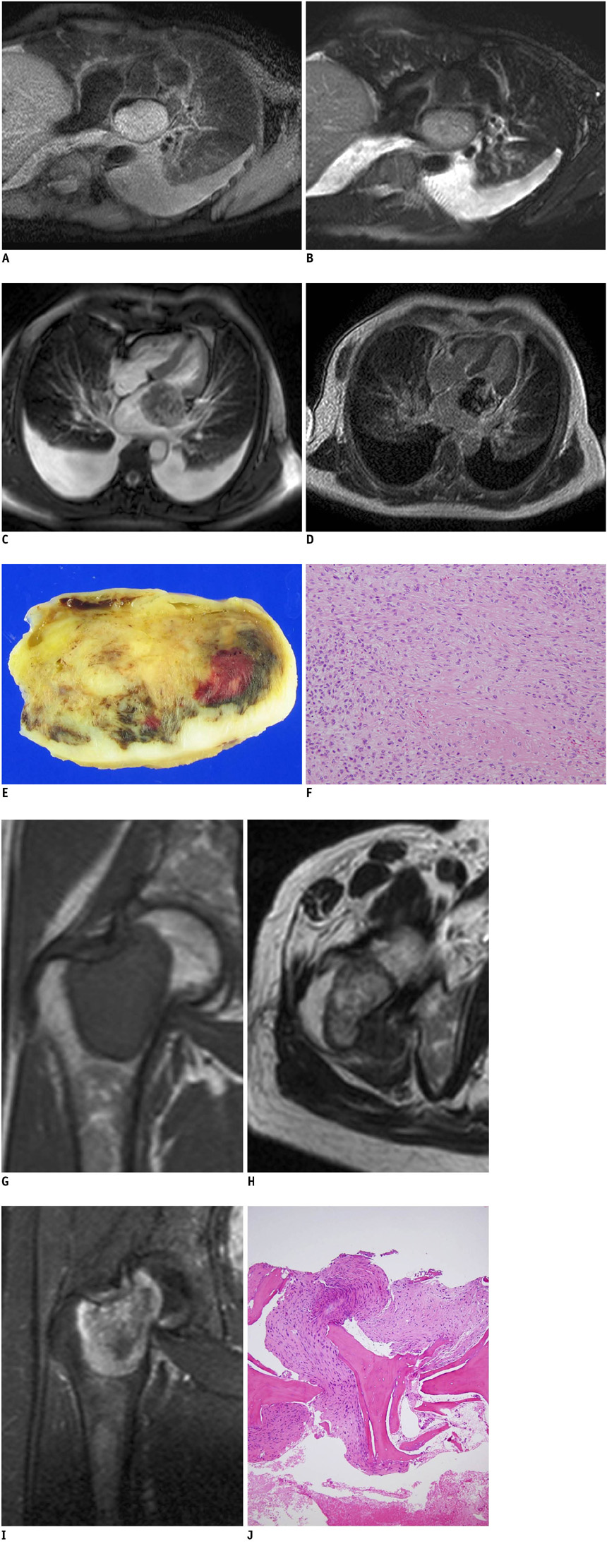
Fig. 1 MRI and histologic finding of cardiac osteosarcoma and its femoral metastasis. A. Axial double inversion-recovery T1-weighted image with fat saturation (TR/TE, 960/8.6 msec) showed well-demarcated mass of mildly heterogeneous and high signal intensity without evidence of invasion into surrounding structures. B, C. Axial triple inversion-recovery T2-weighted image with fat saturation (TR/TE, 960/100 msec) and four chamber view of cine image (3211/1605 msec) showed heterogeneous signal intensity of mass. D. Axial image of delayed contrast-enhanced MRI (TR/TE, 4457/1427 msec) showed heterogeneous enhancement. E. Mass measured about 5.0 × 3.7 × 2.3 cm and had focal hemorrhage and necrosis on gross specimen. F. Photomicrograph showed atypical spindle cells with abundant collagen material and displayed focal immature osteoid production (Hematoxylin & Eosin stain, × 40). G. T1-weighted coronal image (TR/TE, 651/20 msec) showed mass that was isointense to muscle at right femoral neck. Peripheral rim with low signal intensity was seen. H. T2-weighted axial image (TR/TE, 4053/100 msec) showed heterogeneously high signal intensity lesion with peripheral hypointense rim at right femoral neck. I. Gadolinium-enhanced T1-weighted coronal image with fat suppression (TR/TE, 540/17 msec) was performed and lesion showed heterogeneous enhancement within central portion of non-enhancement. J. Atypical spindle cells with abundant collagen material were seen on photomicrograph (Hematoxylin & Eosin stain, × 40).
Reference
-
1. Roberts WC. Primary and secondary neoplasms of the heart. Am J Cardiol. 1997. 80:671–682.2. Sparrow PJ, Kurian JB, Jones TR, Sivananthan MU. MR imaging of cardiac tumors. Radiographics. 2005. 25:1255–1276.3. Araoz PA, Eklund HE, Welch TJ, Breen JF. CT and MR imaging of primary cardiac malignancies. Radiographics. 1999. 19:1421–1434.4. O'Donnell DH, Abbara S, Chaithiraphan V, Yared K, Killeen RP, Cury RC, et al. Cardiac tumors: optimal cardiac MR sequences and spectrum of imaging appearances. AJR Am J Roentgenol. 2009. 193:377–387.5. Takeuchi I, Kawaguchi T, Kimura Y, Kojima J, Shimamura H, Shimizu N, et al. Primary cardiac osteosarcoma in a young man with severe congestive heart failure. Intern Med. 2007. 46:649–651.6. Gomez-Rubin MC, Rios JC, Dobarro D, Sanchez-Recalde A, Bret-Zurita M, Filgueiras D, et al. A recidivant primary cardiac osteosarcoma: the role of bone scans. Cardiovasc Pathol. 2010. 19:55–58.7. Kim EY, Choe YH, Sung K, Park SW, Kim JH, Ko YH. Multidetector CT and MR imaging of cardiac tumors. Korean J Radiol. 2009. 10:164–175.8. Yamagishi M, Yamada N, Kuribayashi S. Images in cardiology: magnetic resonance imaging of cardiac osteosarcoma. Heart. 2001. 85:311.9. Lurito KJ, Martin T, Cordes T. Right atrial primary cardiac osteosarcoma. Pediatr Cardiol. 2002. 23:462–465.10. Vanhoenacker FM, Van de Perre S, Van Marck E, Somville J, Gielen JL, De Schepper AM. Extraskeletal osteosarcoma: report of a case with unusual imaging features and histopathological correlation. Eur J Radiol Extra. 2004. 49:97–102.
- Full Text Links
-
- Actions
-
Cited
- CITED
-
- Close
- Share
-
- Similar articles
-
- MR Imaging Findings of Chondroblastic Osteosarcoma
- Transphyseal extension of osteosarcoma: MRI and pathologic correlation
- MR Imaging of Osteosarcoma: Emphasis on Joint Involvment
- Development of Conventional Osteosarcoma after 13 Years Continuous Disease-free Survival of Periosteal Osteosarcoma
- FDG-PET/CT Complements Bone Scan with Respect to the Detection of Skip Metastasis of Osteosarcoma: A Case Report
