Imaging Findings of Follicular Dendritic Cell Sarcoma: Report of Four Cases
- Affiliations
-
- 1Department of Radiology, Affiliated HuaShan Hospital, Fudan University, 12 Wulumuqi Road, Shanghai 200040, China.
- 2Department of Radiology, Affiliated Cancer Hospital, Fudan University, 270 Dongan Road, Shanghai 200032, China. 081105209@fudan.edu.cn
- 3Department of Pathology, Affiliated Cancer Hospital, Fudan University, 270 Dongan Road, Shanghai 200032, China.
- KMID: 991690
- DOI: http://doi.org/10.3348/kjr.2011.12.1.122
Abstract
- Follicular dendritic cell sarcoma is a rare malignant neoplasm and little is known about its radiological features. We present here four cases of follicular dendritic cell sarcomas and we provide the image characteristics of these tumors to help radiologists recognize this entity when making a diagnosis.
Keyword
MeSH Terms
Figure
-

Fig. 1 Follicular dendritic cell sarcoma in mediastinum in 47-year-old man. A. Unenhanced CT image of thorax reveals well-defined posterior mediastinal mass of homogeneous attenuation (white arrows) with arborizing-pattern of calcification (black arrow). B. Contrast-enhanced CT image shows marked homogeneous enhancement of mass. Note compression of left atrium (black asterisk) and displacement of esophagus (white arrow).
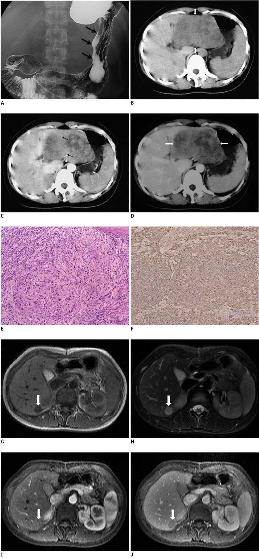
Fig. 2 Follicular dendritic cell sarcoma in upper abdomen in 28-year-old woman. A. Image of air-barium double-contrast study shows broadening of incisura due to extrinsic compression along lesser curvature of stomach (black arrows). Overlying mucosa appears to be intact. B. Unenhanced CT image of upper abdomen shows large heterogeneous mass (white arrows) located between stomach and left lobe of liver. C. Contrast-enhanced CT image during arterial phase shows heterogeneous moderate enhancement of tumor. Note feeding arteries in periphery of tumor (black arrows). D. Portal venous phase image shows heterogeneous moderate contrast enhancement of tumor (white arrows). E. Histopathological appearance reveals that tumor is composed of spindle cells that are arranged in storiform and whorled pattern and these spindle cells are admixed with lymphocytes (Hematoxylin & Eosin stain, × 100). F. Tumor shows positive immunohistochemical staining for CD21 (paraffin immunohistochemical stain, × 100). G. T1-weighted image shows hypointense metastatic nodule in right lobe of liver (white arrow). H. Nodule is hyperintense with hypointense center on T2-weighted image (white arrow). I. T1-weighted arterial-phase contrast-enhanced image shows isointensity of nodule (white arrow) due to homogeneous enhancement. J. T1-weighted portal-phase enhanced image shows heterogeneous, mild hypointensity of nodule (white arrow).
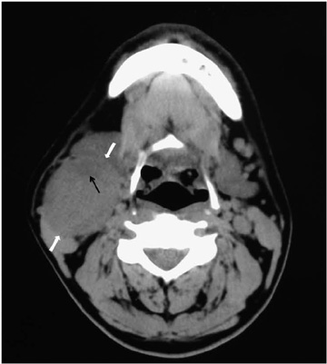
Fig. 3 Follicular dendritic cell sarcoma of cervical lymph node in 38-year-old man. Unenhanced axial CT image reveals well-delineated homogeneous right submandibular mass that compresses adjacent structures (white arrows). Small area of mild hypodensity (black arrow) is present in mass.
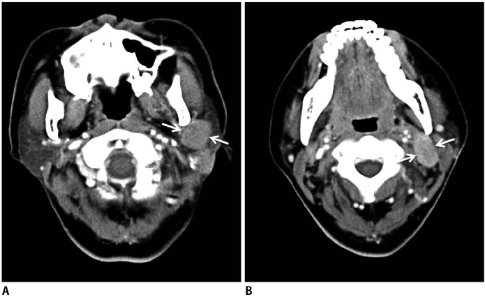
Fig. 4 Follicular dendritic cell sarcoma of cervical lymph nodes in 35-year-old woman. A, B. Enhanced CT images show multiple enlarged lymph nodes with homogenous moderate enhancement (white arrows) in left parotid gland region.
Reference
-
1. Monda L, Warnke R, Rosai J. A primary lymph node malignancy with features suggestive of dendritic reticulum cell differentiation. A report of 4 cases. Am J Pathol. 1986. 122:562–572.2. Weiss LM, Grogan TM, Muller-Hermelink H-K, Stein H, Pura T, Favara B, et al. Swerdlow SH, Campo E, Harris NL, Jaffe ES, Pileri SA, Stein H, editors. Histiocytic and dendritic cell neoplasms. WHO classification of tumours of haematopoietic and lymphoid tissues. 2008. 4th ed. Lyon: IARC Press;286–288.3. Leipsic JA, McAdams HP, Sporn TA. Follicular dendritic cell tumor of the mediastinum. AJR Am J Roentgenol. 2007. 188:W554–W556.4. Kang TW, Lee SJ, Song HJ. Follicular dendritic cell sarcoma of the abdomen: the imaging findings. Korean J Radiol. 2010. 11:239–243.5. Shia J, Chen W, Tang LH, Carlson DL, Qin J, Guillem JG, et al. Extranodal follicular dendritic cell sarcoma: clinical, pathologic, and histogenetic characteristics of an underrecognized disease entity. Virchows Arch. 2006. 449:148–158.6. Chan JK, Fletcher CD, Nayler SJ, Cooper K. Follicular dendritic cell sarcoma. Clinicopathologic analysis of 17 cases suggesting a malignant potential higher than currently recognized. Cancer. 1997. 79:294–313.7. Perez-Ordonez B, Erlandson RA, Rosai J. Follicular dendritic cell tumor: report of 13 additional cases of a distinctive entity. Am J Surg Pathol. 1996. 20:944–955.8. Shek TW, Liu CL, Peh WC, Fan ST, Ng IO. Intra-abdominal follicular dendritic cell tumour: a rare tumour in need of recognition. Histopathology. 1998. 33:465–470.9. Díaz de Liaño A, Garde C, Artieda C, Yárnoz C, Flores L, Ortiz H. Intra-abdominal follicular dendritic cell sarcoma. Clin Transl Oncol. 2006. 8:837–838.10. Jiang L, Admirand JH, Moran C, Ford RJ, Bueso-Ramos CE. Mediastinal follicular dendritic cell sarcoma involving bone marrow: a case report and review of the literature. Ann Diagn Pathol. 2006. 10:357–362.11. Ceresoli GL, Zucchinelli P, Ponzoni M, Gregorc V, Bencardino K, Paties CT. Mediastinal follicular dendritic cell sarcoma. Haematologica. 2003. 88:ECR04.12. Ko SF, Hsieh MJ, Ng SH, Lin JW, Wan YL, Lee TY, et al. Imaging spectrum of Castlemans' disease. AJR Am J Roentgenol. 2004. 182:769–775.13. Chang KC, Jin YT, Chen FF, Su IJ. Follicular dendritic cell sarcoma of the colon mimicking stromal tumour. Histopathology. 2001. 38:25–29.14. Bai LY, Kwang WK, Chiang IP, Chen PM. Follicular dendritic cell tumor of the liver associated with Epstein-Barr virus. Jpn J Clin Oncol. 2006. 36:249–253.15. O'Malley DP. Diagnosis: follicular dendritic cell tumor mimicking GI stromal tumor. 2004. In: http://socforheme.org/case-nov-04.htm.16. Chen TC, Kuo TT, Ng KF. Follicular dendritic cell tumor of the liver: a clinicopathologic and Epstein-Barr virus study of two cases. Mod Pathol. 2001. 14:354–360.
- Full Text Links
-
- Actions
-
Cited
- CITED
-
- Close
- Share
-
- Similar articles
-
- Follicular Dendritic Cell Sarcoma of the Abdomen: the Imaging Findings
- Recurrent Follicular Dendritic Cell Sarcoma of the Parotid Gland Imaged with 18F-FDG PET/CT
- Follicular Dendritic Cell Sarcoma of the Omentum: Multidetector Computed Tomography Findings
- Follicular Dendritic Cell Sarcoma of the Tonsil
- Follicular dendritic cell sarcoma: Rare presentation of incidental large hepatic mass
