Solitary Necrotic Nodules of the Liver Mimicking Hepatic Metastasis: Report of Two Cases
- Affiliations
-
- 1Department of Radiology, Wonkwang University School of Medicine, Iksan, Chunbuk, Korea. yoonkh@wmc.wonkwang.ac.kr
- KMID: 877078
- DOI: http://doi.org/10.3348/kjr.2000.1.3.165
Abstract
- We present two cases of solitary necrotic nodules of the liver which on radiologic images mimicked hepatic metastasis. Solitary necrotic nodule of the liver is a rare but benign entity which histopathologically consists of an outer fibrotic cap-sule with inflammatory cells and a central core of amorphous necrotic material. The lesion was seen on contrast-enhanced CT as an ovoid-shaped hypoattenu-ating nodule; on CT during hepatic arteriography as enhancing nodule; on intra-operative US as a target-appearing hypoechoic nodule; on T2WI as a hyperinten-sity nodule, and on dynamic MR as a subtle peripheral enhancing nodule. Although the radiologic features are not specific, solitary necrotic nodule of the liver should be included in the differential diagnosis of hepatic metastasis.
Keyword
MeSH Terms
Figure
-

Fig. 1 A 54-year-old woman with gallbladder cancer and solitary necrotic nodule of the liver. A. CTAP shows an ovoid-shaped area of portal perfusion defect (arrows), 1.0 cm in diameter, in the right hepatic lobe. B. CTHA shows a highly enhancing nodular lesion (arrows) in the same area. C. Intraoperative US shows an additional target-appearing hypoechoic nodule (arrows), 0.5 cm in diameter, in the same segment of the liver. D. Photomicrograph shows central amorphous necrosis (arrows) surrounded by a fibrotic capsule, with infiltration by inflammatory cells (arrowheads) (hematoxylin-eosin stain, ×13).
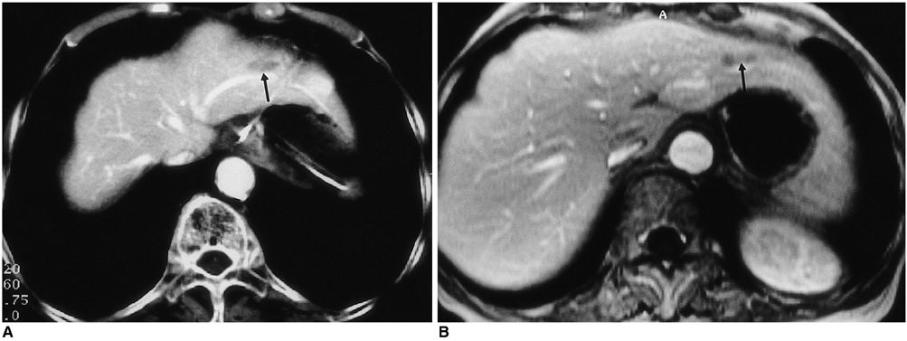
Fig. 2 A 72-year-old man who six years earlier had undergone subtotal gastrectomy due to gastric cancer. A. Contrast-enhanced CT shows an elliptical hypoattenuating nodule (arrow) in the lateral segment of the liver. B. Contrast-enhanced T1-weighted MR image shows a subtle peripheral enhancing nodule (arrow) at the same site.
Reference
-
1. Shepherd NA, Lee G. Solitary necrotic nodules of the liver stimulating hepatic metastases. J Clin Pathol. 1983. 36:1181–1183.2. Alfieri S, Carriero C, Doglietto GB, Pacelli F, Crucitti F. Solitary necrotic nodule of the liver: diagnosis and treatment. Hepatogastroenterology. 1997. 44:1210–1211.3. Tsui WMS, Yuen RWS, Chow LTC, Tse CCH. Solitary necrotic nodule of the liver: parasitic origin? J Clin Pathol. 1992. 45:975–978.4. Berry CL. Solitary necrotic nodule of the liver: a probable pathogenesis. J Clin Pathol. 1985. 38:1278–1280.5. Sundaresan M, Lyons B, Akosa AB. 'Solitary' necrotic nodules of the liver: an etiology reaffirmed. Gut. 1991. 32:1378–1380.6. Soyer P, Levesque M, Elias D, Zeitoun G, Roche A. Preoperative assessment of resectability of hepatic metastases from colon carcinoma: CT portography vs sonography and dyanamic CT. AJR. 1992. 159:741–744.7. Hagspiel KD, Neidl KFW, Eichenberger AC, Weder W, Marincek B. Detection of liver metastases: comparison of superparamagnetic iron oxide-enhanced and unenhanced MR imaging at 1.5 T with dynamic CT, intraoperative US, and percutaneous US. Radiology. 1995. 196:471–478.8. Yoon K-H, Ha HK, Lee JS, et al. Inflammatory pseudotumor of the liver in patients with recurrent pyogenic cholangitis: CT-histopathologic correlation. Radiology. 1999. 211:373–379.9. Won JH, Kim M-J, Kim BM, et al. Focal eosinophilic infiltration of the liver: a mimick of hepatic metastasis. Abdom Imaging. 1999. 24:369–372.10. Lee WJ, Lim HK, Lim JH, Kim SH, Choi SH, Lee SJ. Foci of eosinophil-related necrosis in the liver: imaging findings and correlation with eosinophilia. AJR. 1999. 172:1255–1261.
- Full Text Links
-
- Actions
-
Cited
- CITED
-
- Close
- Share
-
- Similar articles
-
- Solitary Necrotic Nodule of the Liver Mimicking Metastasis in Patient with Early Gastric Cancer: 3T MRI and PET/CT Findings
- Hepatic Fascioliasis Mimicking Metastatic Tumor
- A Large Solitary Liver Metastasis of Thymoma
- Solitary Extrahepatic Intraabdominal Metastasis from Hepatocellular Carcinoma after Liver Transplantation
- Malignant Hepatic Solitary Fibrous Tumor
