Teratoma with Malignant Transformation in the Anterior Mediastinum: A Case Report
- Affiliations
-
- 1Department of Radiology, St. Mary's Hospital, College of Medicine, The Catholic University of Korea, Seoul, Korea. jijung@cmc.cuk.ac.kr
- KMID: 877077
- DOI: http://doi.org/10.3348/kjr.2000.1.3.162
Abstract
- Malignant transformation of teratoma in the anterior mediastinum is rare; the mass usually has a long history and is seen in older patients. We report a case of teratoma with malignant transformation in the anterior mediastinum, complicated by rupture. CT revealed a lobulated, inhomogeneous cystic mass with a fat com-ponent and wall calcifications. The lateral wall was disrupted and consolidation in the adjacent left upper lobe was noted, suggesting rupture. A heterogeneously enhanced solid portion, obliterating the fat plane between the mass and the great vessels was present in the medial aspect of the mass, and pathologic examina-tion demonstrated the presence of adenocarcinoma.
MeSH Terms
Figure
-
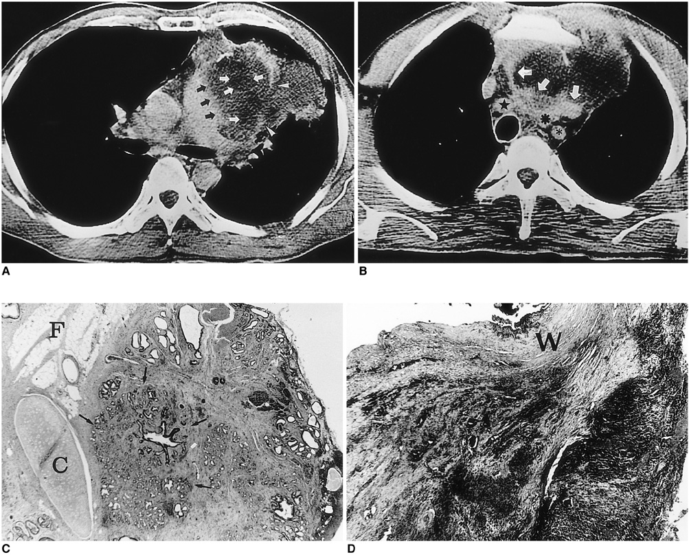
Fig. 1 A-49-year-old man and teratoma and malignant transformation. A. Contrast-enhanced CT scan indicates the presence in the mediastinum of a lobulated low attenuated mass containing a fat component (HU =-45) (white arrows) and wall calcifications. The lateral wall of the mass is focally disrupted and consolidation with the fat component in the adjacent left upper lobe is noted (arrowheads). A heterogeneously enhanced solid portion is observed in the medial aspect of the mass, and this obliterates the fat plane between the mass and the main pulmonary artery (black arrows). B. CT scan obtained at the origin of the great vessels shows an inhomogeneously enhanced solid portion which invades the mediastinal great vessels (white arrows) and was pathologically proven to be adenocarcinoma (★right bracheocephalic artery, ✱left common carotid artery, *left subclavian artery). C. Light microscopic examination demonstrates that part of the resected mass is composed of mature fat (F), chrondroid cartilage (C), and glandular tissue (arrows) (×20, Hemtoxyline-eosin stain). D. Light microscopic examination demonstrates the medial part of the mass, in which poorly differentiated adenocarcinoma crosses the fibrous outer wall (W) (×20, Hematoxyline-eosin stain).
Reference
-
1. Ulbright TM, Loehrer PJ, Roth LM, Einhorn LH, Williams SD, Clark SA. The development of non-germ cell malignancies within germ cell tumors. A clinicopathologic study of 11 cases. Cancer. 1984. 54:1824–1833.2. Ahmed T, Bosl GJ, Hajdu SI. Teratoma with malignant transformation in germ cell tumors in men. Cancer. 1985. 56:860–863.3. Morinaga S, Nomori H, Kobayashi R, Atsumi Y. Well-differentiated adenocarcinoma arising from mature cystic teratoma of the mediastinum (teratoma with malignant transformation): Report of a surgical case. Am J Clin Pathol. 1994. 101:531–534.4. Sasaka K, Kurihara Y, Nakajima Y, et al. Spontaneous rupture: a complication of benign mature teratomas of the mediastinum. AJR. 1998. 170:323–328.5. Chadha S, Schaberg A. Malignant transformation in benign cystic teratomas: dermoids of the ovary. Eur J Obstet Gynecol Reprod Biol. 1988. 19:329–338.6. Curling OM, Potsides PN, Hudson CN. Malignant change in benign cystic teratoma of the ovary. Br J Obstet Gynecol. 1979. 86:399–402.7. Knapp RH, Hurt RD, Payne WS, et al. Malignant germ cell tumors of the mediastinum. J Thorac Cardiovasc Surg. 1985. 89:82–89.8. Moeller KH, Rosado-de-Christenson ML, Templeton PA. Mediastinal mature teratoma: imaging features. AJR. 1997. 169:985–990.9. Choi S, Lee JS, Song KS, Lim T. Mediastinal teratoma: CT differentiation of ruptured and unruptured tumors. AJR. 1998. 171:591–594.
- Full Text Links
-
- Actions
-
Cited
- CITED
-
- Close
- Share
-
- Similar articles
-
- Adenocarcinoma Arising in benign Teratoma of Mediastinum: A case report
- Naturally Occurring Mediastinal Teratoma with Malignant Transformation in an Adult Male
- Malignant Teratoma of Prostatic Gland
- Rapidly Growing Mature Teratoma in the Anterior Mediastinum: Case Report
- Teratoma of the Pleura: A Case Report
