Korean J Ophthalmol.
2009 Dec;23(4):329-331. 10.3341/kjo.2009.23.4.329.
A Case of Pseudo-Duane's Retraction Syndrome With Old Medial Orbital Wall Fracture
- Affiliations
-
- 1Department of Ophthalmology, Daegu Catholic University College of Medicine, Daegu, Korea. kimsy@cu.ac.kr
- KMID: 754776
- DOI: http://doi.org/10.3341/kjo.2009.23.4.329
Abstract
- We report a case of pseudo-Duane's retraction syndrome with entrapment of the medial rectus muscle in an old medial orbital wall fracture presenting identical clinical symptoms as Duane's retraction syndrome. A 15-year-old boy presented with persistent limited right eye movement since a young age. Examination showed marked limited abduction, mildly limited adduction, and globe retraction accompanied by narrowing of the palpebral fissure during attempted adduction in the right eye. He showed a right esotropia of 16 prism diopters and his head turned slightly to the right. A slight enophthalmos was noted in his right eye. A computed tomography scan demonstrated entrapment of the medial rectus muscle and surrounding tissues in an old medial orbital wall fracture. A forced duction test revealed a marked restriction of abduction in the right eye. A 5 mm recession of the right medial rectus muscle was performed. Postoperatively, the patient's head turn and esotropia in the primary position were successfully corrected, but there was still some limitations to his ocular movement. The importance of several tests such as the forced duction test and an imaging study should be emphasized in making a diagnosis for limitation of eye movement.
Keyword
MeSH Terms
Figure
-
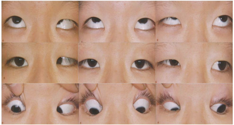
Fig. 1 Preoperative photographs show markedly limited abduction, mildly limited adduction, and narrowing of the palpebral fissure on adduction of the right eye.

Fig. 2 Side view of the right eye shows. (A) A slight enophthalmos in the primary position. (B) Globe retraction upon attempted adduction.

Fig. 3 Preoperative orbital CT scan shows an old fracture of the right medial orbital wall with entrapment of the right medical rectus muscle. (A) Axial view. (B) Corneal view.
Reference
-
1. Duane TD, Schatz NJ, Caputo AR. Pseudo-Duane's retraction syndrome. Trans Am Ophthalmol Soc. 1976. 74:122–132.2. DeRespinis PA, Caputo AR, Wagner RS, Guo S. Duane's retraction syndrome. Surv Ophthalmol. 1993. 38:257–288.3. Gittinger JW Jr, Hughes JP, Suran EL. Medial orbital wall blow-out fracture producing an acquired retraction syndrome. J Clin Neuroophthalmol. 1986. 6:153–156.4. Chatterjee PK, Bhunia J, Bhattacharyya I. Bilateral inverse Duane's retraction syndrome: a case report. Indian J Ophthalmol. 1991. 39:183–185.5. Khan AO. Inverse globe retraction syndrome complicating recurrent pterygium. Br J Ophthalmol. 2005. 89:640–641.6. Murthy R. Duane's retraction syndrome following myocysticercosis. Indian J Ophthalmol. 2008. 56:89–90.7. Jo KI, Jo YH, Rho YB, et al. A case of inverse Duane's retraction syndrome. J Korean Ophthalmol Soc. 1980. 21:615–619.8. Lew H, Lee JB, Kim HS, Han SH. A case of congenital inverse Duane's retraction syndrome. Yonsei Med J. 2000. 41:155–158.9. Khan AO. Bilateral inverse globe retraction (Duane's) syndrome. Indian J Ophthalmol. 2007. 55:388–389.
- Full Text Links
-
- Actions
-
Cited
- CITED
-
- Close
- Share
-
- Similar articles
-
- A case of congenital inverse Duane's retraction syndrome
- Two Cases of Duane's Retraction Syndrome
- The Direct Medial Canthal Approach in the Repair of Blowout Fracture of the Medial Orbital Wall
- Surgical Treatment of the Upshoot and Downshoot in Duane's Retraction Syndrome
- A Case of Inverse Duane's Retraction Syndrome
