A Case of Photic Retinal Injury Associated with Exposure to Plasma Arc Welding
- Affiliations
-
- 1Department of Ophthalmology, Wonju Christian Hospital, Yonsei University Wonju College of Medicine, Wonju-city, Kangwon-do, Korea. eyesj@yonsei.ac.kr
- KMID: 754593
- DOI: http://doi.org/10.3341/kjo.2006.20.4.250
Abstract
- PURPOSE: To report of photic retinopathy induced by plasma arc welding, and the OCT (optical coherence tomography) results of damaged retinal lesions. METHODS: We describe a case report of a 37-year-old male, working in the steel industry, who presented with central scotoma in both eyes. RESULTS: On his first visit, one day after performing plasma arc welding with protective gear at work, his best corrected vision was 0.7 for both eyes. Ophthalmic examination of the fundus showed a round yellow lesion with an approximate size of 300 micrometers superonasal to the fovea of both eyes. On his next visit, one month later, his vision had recovered to 1.0, his symptoms had improved, and the ophthalmoscopic examination of the fundus revealed that the round yellow spots had disappeared from both eyes. CONCLUSIONS: To our knowledge, this is the first report of photic retinopathy induced by plasma arc welding, and the OCT (optical coherence tomography) results of damaged retinal lesions have not previously been reported. For these reasons, we report this case.
Keyword
MeSH Terms
Figure
-
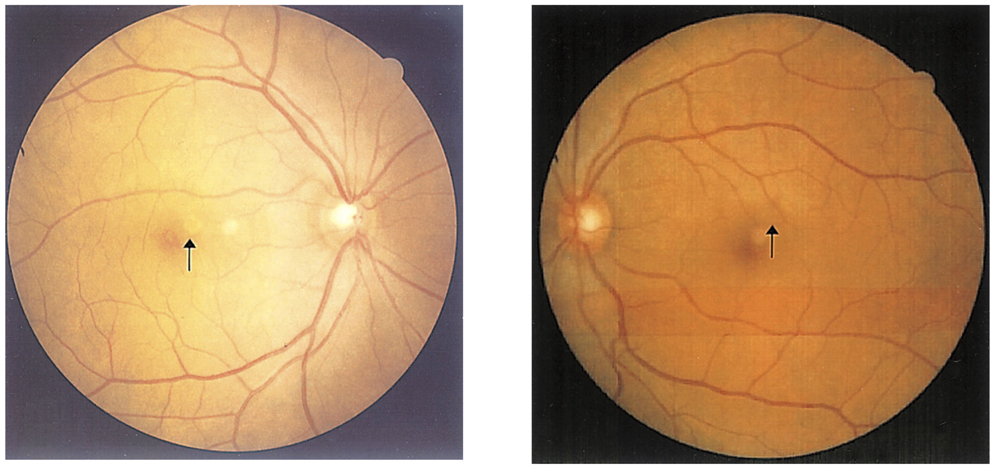
Fig. 1 Fundus photograph shows a round yellowish lesion at the parafoveal area (both eyes).
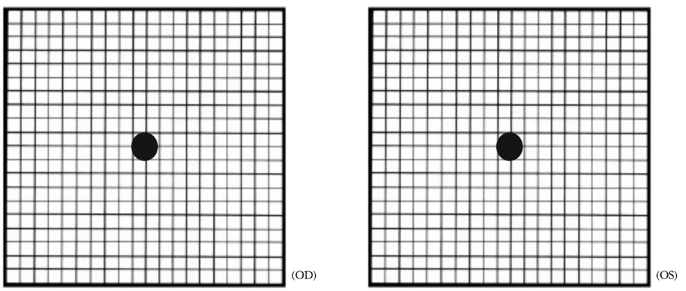
Fig. 2 Amsler grid test shows a round scotoma in the central region (both eyes).
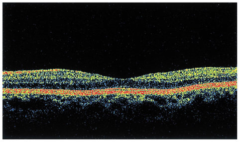
Fig. 3-1 The OCT shows the normal finding of the cross-sectional retinal image (right eye).
Fig. 3-2 The OCT shows the normal finding of the cross0sectional retinal image (left eye).
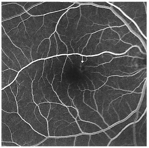
Fig. 4 Ring-shaped paracentral hyperfluorescence appears simultaneously with a central halo in the early phase (white arrow, right eye).

Fig. 5 The dark center of the lesion is surrounded by progressively increasing hyperfluorescence in the late phase (white arrow, right eye).
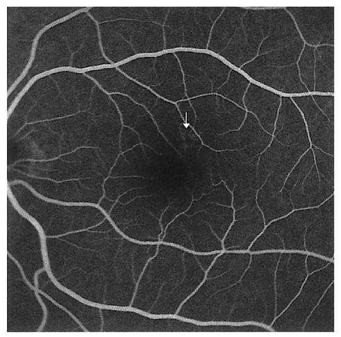
Fig. 6 Ring-shaped paracentral hyperfluorescence appears simultaneously with a central halo in the early phase (white arrow, left eye).
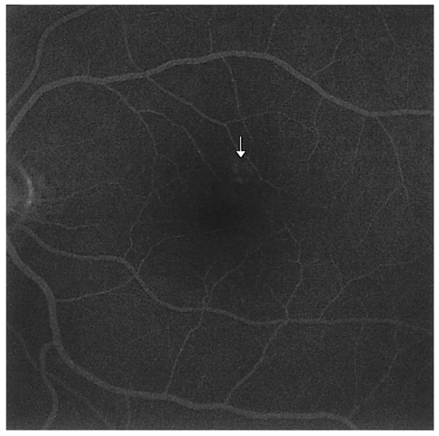
Fig. 7 The dark center of the lesion is surrounded by progressively increasing hyperfluorescence in the late phase (white arrow, left eye).
Cited by 1 articles
-
A Case of Maculopathy from Handheld Green Laser Pointer
Young Jun Kim, In Young Chung, Seong Jae Kim, Jong Moon Park, Yong Seop Han
J Korean Ophthalmol Soc. 2015;56(3):447-451. doi: 10.3341/jkos.2015.56.3.447.
Reference
-
1. Brown J, Planck S, Meshul C, et al. Ultraviolet irradiation induces the production of multiple cytokines by human corneal cells. Invest Ophthalmol Vis Sci. 1997. 38:2483–2490.2. Arend O, Aral H, Reim M, et al. Welders maculopathy despite using protective lenses. Retina. 1996. 16:257–259.3. Freeman J, Gombos GM. Fluorescein fundus angiography in self-induced solar retinopathy. A case report. Can J Ophthalmol. 1971. 6:124–127.4. Ham WT Jr, Mueller HA, Rufflo JJ Jr, et al. Basic mechanisms underlying the production of photochemical lesions in the mammalian retina. Curr Eye Res. 1984. 3:165–174.5. Lawwill T. Three major pathologic processes caused by light in the primate retina: a search for mechanisms. Trans Am Ophthalmol Soc. 1982. 80:517–579.6. Naidoff MA, Slinkey DH. Retinal injury from a welding arc. Am J Ophthalmol. 1974. 77:663–668.7. Romanchuk KG, Pollak V, Schneider RJ. Retinal burn from a welding arc. Can J Ophthalmol. 1978. 13:120–122.8. Uniat L, Olk RJ, Hanish SJ. Welding arc maculopathy. Am J Ophthalmol. 1986. 102:394–395.9. Shahriari HA, Salari AM. Preventive effects of Vitamin A and Aspirin on the UV light-induced retinopathy in an animal model. 98134 Zahedan, Iran: Zahedan University of Medical sciences.
- Full Text Links
-
- Actions
-
Cited
- CITED
-
- Close
- Share
-
- Similar articles
-
- Long-term Follow-up Results of Patients with Welding-arc Maculopathy Assessed Using Spectral Domain Optical Coherence Tomography
- Photic Retinal Injury Induced by Endoillumination during Vitrectomy
- Study on the Pulmonary Function in Welding Fume Exposed Workers
- Performance of Neurobehavioral Tests Among Welders Exposed to Manganese
- Industrial Photophthalmia
