Intraventricular Hemorrhage Caused by Lateral Ventricular Meningioma: A Case Report
- KMID: 754112
- DOI: http://doi.org/10.3348/kjr.2001.2.2.105
Abstract
- Meningiomas causing intracranial hemorrhage are rare, and hemorrhage from a lateral ventricular meningioma seems to be even rarer. We report a case of trigonal meningioma in a 43-year-old woman who presented with intraventricular hemorrhage, and describe the CT, MRI and angiographic findings.
Keyword
MeSH Terms
Figure
-
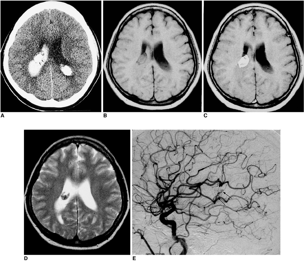
Fig. 1 Images from the case of a 43-year-old woman with sudden headache. A. Noncontrast axial CT scan obtained on day 1 shows dense calcified masses (arrows) in the trigones of both lateral ventricles, combined with intraventricular hemorrhage in the right lateral ventricle. The right lateral ventricular mass extends to the body of the lateral ventricle. B. Axial T1-weighted image shows that the right lateral ventricular mass is almost isointense to brain parenchyma. Intraventricular hemorrhage is also present. C. Axial contrast-enhanced T1-weighted image(500/16) shows intense enhancement of the right lateral ventricular mass. The left lateral ventricular mass (not shown) exhibited inhomogeneous enhancement. D. Axial T2-weighted image(2500/80) reveals the presence of an inhomogeneous mass in the right lateral ventricle. The low signal intensity suggets the presence of calcification and hemorrhage. E. Angiogram of the right internal carotid artery obtained on day 3 demonstrates a hypervascular mass fed from the right anterior choroidal artery (arrows).
Reference
-
1. Marti-Fabregas J, Piles S, Guardia E, Marti-Vilalta JL. Spontaneous primary intraventricular hemorrhage: clinical data, etiology and outcome. J Neurol. 1999. 246:246–291.2. Kendall B, Grosswasser IR, Valentine A. Diagnosis of masses presenting within the ventricles on computed tomography. Neuroradiology. 1983. 25:11–22.3. Lang I, Jackson A, Strang FA. Intraventricular hemorrhage caused by intraventricular meningioma: CT appearance. AJNR. 1995. 16:1378–1381.4. Murai Y, Yochida D, Ikeda Y, et al. Spontaneous intraventricular hemorrhage caused by lateral ventricular meningioma; A case report. Neurol Med Chir (Tokyo). 1996. 36:586–558.5. Morrison G, Sobel DF, Kelley WM, Norman D. Intraventricular mass lesions. Radiology. 1984. 153:435–442.6. Guidetti B, Delfini R, Gagliardi FM, Vagnozzi R. Meningiomas of the lateral ventricles. Surg Neurol. 1985. 24:364–370.7. Jelinek J, Smirniotopoulos JG. Lateral ventricular neoplasm of the brain: differentiatial diagnosis based on clinical, CT and MR findings. Am J Neuroradiol. 1990. 11:567–574.8. Smith VR, Stein PS, MacCarty CS. Subarachnoid hemorrhage due to lateral ventricular meningioma. Surg Neurol. 1975. 4:241–243.
- Full Text Links
-
- Actions
-
Cited
- CITED
-
- Close
- Share
-
- Similar articles
-
- A Case of Intraventricular Meningioma Acompanied by Intraventricular Hematoma and Subarachnoid Hemorrhage: Case Report
- Lateral Ventricular Meningioma Presenting with Intraventricular Hemorrhage
- A Case of Giant Intraventricular Meningioma
- Intraventricular Malignant Meningioma with CSF-Disseminated Spinal Metastasis : Case Report and Literature Review
- Change of the Ventricular System Following Intraventricular Injection of the Blood
