Fetal Pericallosal Lipoma: US and MR Findings
- Affiliations
-
- 1Department of Radiology, Sejong Heart Institute, Sejong General Hospital, Pucheon, Korea. fetalus@naver.com
- 2Department of Obstetrics, Sejong Heart Institute, Sejong General Hospital, Pucheon, Korea.
- 3Department of Gynecology, Sejong Heart Institute, Sejong General Hospital, Pucheon, Korea.
- KMID: 754075
- DOI: http://doi.org/10.3348/kjr.2002.3.2.140
Abstract
- We report a case of fetal pericallosal lipoma occurring at the anterior interhemispheric fissure and associated with agenesis of the corpus callosum. During targeted prenatal ultrasonography at 26 weeks' gestation, the lesion was seen as a highly echogenic mass. MR imaging performed at 35 weeks' gestation and during the postnatal period revealed a pericallosal fatty mass and agenesis of the corpus callosum.
MeSH Terms
Figure
-

Fig. 1 Tubulonodular type of pericallosal lipoma in utero. A. Sonogram at 26 weeks' gestation. A hyperechoic mass is seen within the anterior interhemispheric fissure (arrow). The occipital horns of the lateral ventricles are slightly dilated, though the frontal horns are not dilated and are more laterally positioned. The cavum septi pellucidi is not visualized. B-D. MR images at 35 weeks' gestation. The mass shows high signal intensity at T1-weighted axial imaging (arrow) (B) and intermediate signal intensity at T2-weighted axial imaging (arrow) (C). A fat-suppressed T1-weighted MR image (D) shows complete cancellation of the signal intensity of the mass. MR images also demonstrate colpocephaly.
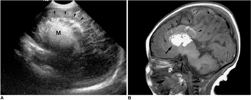
Fig. 2 Tubulonodular-type pericallosal lipoma, as seen at postnatal follow-up imaging. A. Sagittal neurosonogram at the second day shows a hyperechoic midline mass (M), with scattered echogenic spots (arrowheads) in the cingulate sulcus. B. T1-weighted sagittal MR image demonstrates a high-intensity bulky mass in the midline of the anterior callosal area (arrow), with scattered fat globules in the cingulate sulcus (arrowheads). The corpus callosum is invisible. The cerebral gyri extend in a radial pattern from the lateral ventricles, without the normal curve of the cingulate gyrus.
Reference
-
1. Bork MD, Smeltzer JS, Egan JF, Rodis JF, DiMario FJ Jr, Campbell WA. Prenatal diagnosis of intracranial lipoma associated with agenesis of the corpus callosum. Obstet Gynecol. 1996. 87:845–848.2. Ickowitz V, Eurin D, Rypens F, et al. Prenatal diagnosis and postnatal follow-up of pericallosal lipoma: report of seven new cases. AJNR. 2001. 22:767–772.3. Demaerel P, Van de Gaer P, Wilms G, Baert AL. Interhemispheric lipoma with variable callosal dysgenesis: relationship between embryology, morphology, and symptomatology. Eur Radiol. 1996. 6:904–909.4. Multz MA, Koenigsberg M, Lantos G. US case of the day: lipoma and hypogenesis of the corpus callosum. RadioGraphics. 1996. 16:1227–1230.5. Kazner E, Stochdorph O, Wende S, Grumme T. Intracranial lipoma: diagnostic and therapeutic considerations. J Neurosurg. 1980. 52:234–245.6. Truwit CL, Barkovich AJ. Pathogenesis of intracranial lipoma: an MR study in 42 patients. AJR. 1990. 155:855–865.7. Lee SH, Cho JY, Song MJ, et al. Prenatal ultrasound findings of fetal neoplasms. Korean J Radiol. 2002. 3:64–73.8. Jeanty P, Zaleski W, Fleischer AC. Prenatal sonographic diagnosis of lipoma of the corpus callosum in a fetus with Goldenhar syndrome. Am J Perinatol. 1991. 8:89–90.9. Beltinger C, Saule H. Imaging of lipoma of the corpus callosum and intracranial dermoids in the Goldenhar syndrome. Pediatr Radiol. 1989. 18:72–73.10. Tahmouresie A, Kroll G, Shucart W. Lipoma of the corpus callosum. Surg Neurol. 1979. 11:31–34.
