Fetal Musculoskeletal Malformations with a Poor Outcome: Ultrasonographic, Pathologic, and Radiographic Findings
- Affiliations
-
- 1Department of Radiology, Samsung Cheil Hospital, Sungkyunkwan University School of Medicine, Seoul, Korea. radjycho@samsung.co.kr
- 2Department of Radiology, Seoul National University College of Medicine, Seoul, Korea.
- KMID: 754070
- DOI: http://doi.org/10.3348/kjr.2002.3.2.113
Abstract
- The early and accurate antenatal diagnosis of fetal musculoskeletal malfomations with a poor outcome has important implications for the management of a pregnancy. Careful ultrasonographic examination of a fetus helps detect such anomalies, and a number of characteristic features may suggest possible differential diagnoses. During the last five years, we have encountered 39 cases of such anomalies, and the typical prenatal ultrasonographic and pathologic findings of a number of those are described in this article.
MeSH Terms
Figure
-

Fig. 1 Thanatophoric dysplasia in a 21-week fetus. A. Ultrasonogram demonstrates a cloverleaf-like skull. B. Rhizomelic micromelia with bowing of the humerus is apparent (arrows). The skin appears thick because of extreme redundancy. C. The normal abdomen (arrows) is protuberant compared with the small thorax. D. Postmortem radiograph shows generalized severe micromelia and a constricted thorax. Bowing is more apparent in the lower extremities.

Fig. 2 Osteogenesis imperfecta type II in a 22-week fetus. A. Axial image of the fetal head shows decreased echogenicity of the calvaria, through which transmission of the ultrasound beam is abnormally increased. B. Ultrasonogram depicts severe micromelia and deformity secondary to fractures (open arrow). C. Axial image shows multiple rib fractures (arrows), with collapse of the thoracic cage. D. Postmortem radiograph show decreased mineralization and severe micromelia, with innumerable fractures.
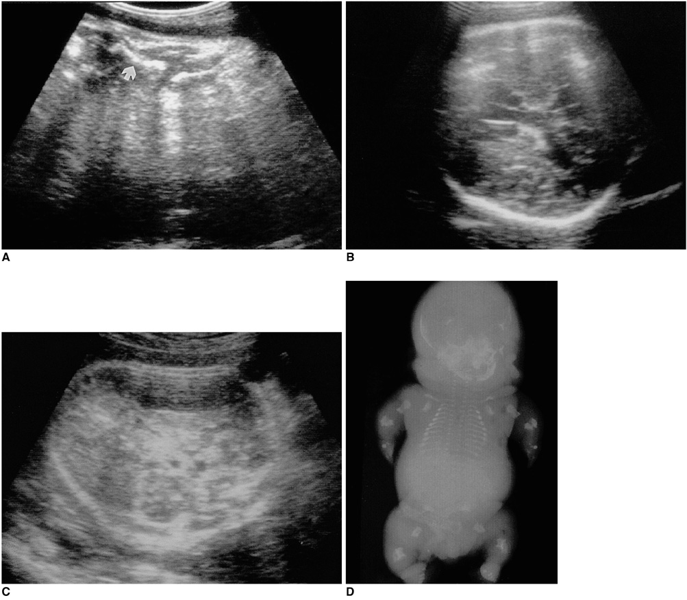
Fig. 3 Achondrogenesis in a 35-week fetus. A. Ultrasonogram shows profound limb shortening (arrow). B, C. Axial images of the fetal head demonstrate decreased calvarial ossification, with increased ultrasonic through-transmission. The fetal head is compressed by the transducer. D. Postmortem radiograph shows extremely short limbs, a large head, and the absence of ossification in the ischia, pubis, vertebral body, and calvarium. The head is disproportionately enlarged and the thorax is small.
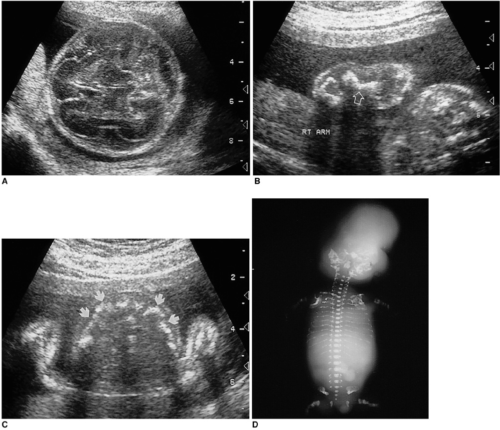
Fig. 4 Short rib dysplasia without polydactyly in a 23-week fetus. A. Ultrasonogram shows short ribs (arrows) and micromelia of the forearm (thin arrow). B. The abdomen is protuberant, and the kidney shows increased echogenicity (arrows), suggesting renal dysplasia. C, D. Postmortem radiograph and photograph depict extremely short ribs, with a narrow chest and protuberant abdomen.
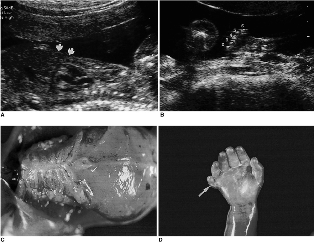
Fig. 5 Short-rib dysplasia with polydactyly in a 21-week fetus. A. Sagittal image shows a narrow thorax and relatively protuberant abdomen (arrows). B. There is an extra digit (6) lateral to the fifth finger. C, D. Autopsy photographs show a narrow thorax with short ribs, a protuberant abdomen, micromelia, and postaxial polydactyly (arrow).
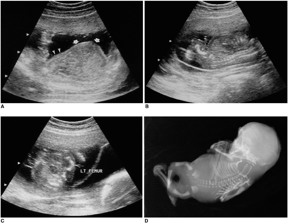
Fig. 6 Camptomelic dysplasia in a 20-week fetus. A. Prenatal ultrasonogram depicts a moderately small thorax (arrowheads) with a protuberant abdomen (arrows). B, C. The femurs (arrow) are short and bowed, and anterolaterally convex. D. Postmortem radiograph demonstrates bowing of the femur and severe clubfoot deformity. The scapula is hypoplastic, but the humerus is relatively preserved.

Fig. 7 Chondrodysplasia punctata in a 35-week fetus. A, B. Prenatal ultrasonograms depict stippled ossification at the proximal epiphyses (arrows) of the femur and humerus. C. There is no abnormal ossification of the distal epiphysis. D. Postnatal radiograph shows stippled ossifications (open arrows) of the proximal femoral epiphyses.
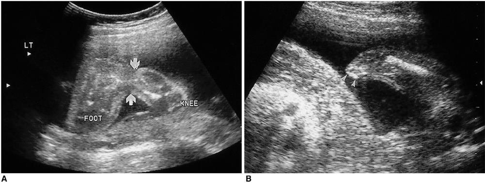
Fig. 8 Amniotic band syndrome in 23-week (A) and 22-week (B) fetuses. A. There is focal constriction (arrows) at the left ankle, with severe lymphedema of the foot. B. The upper extremity is amputated at the proximal forearm (arrowheads).
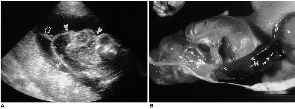
Fig. 9 Amniotic band syndrome with exencephaly and extensive body wall defect. A. Ultrasonogram shows that the calvaria is absent, giving rise to exencephaly (arrows). Note the presence of an adherent amniotic band (open arrow) on the fetal head. B. Postmortem photograph shows exencephaly and asymmetrical body-wall defect, with a herniated heart (H) and abdominal contents.
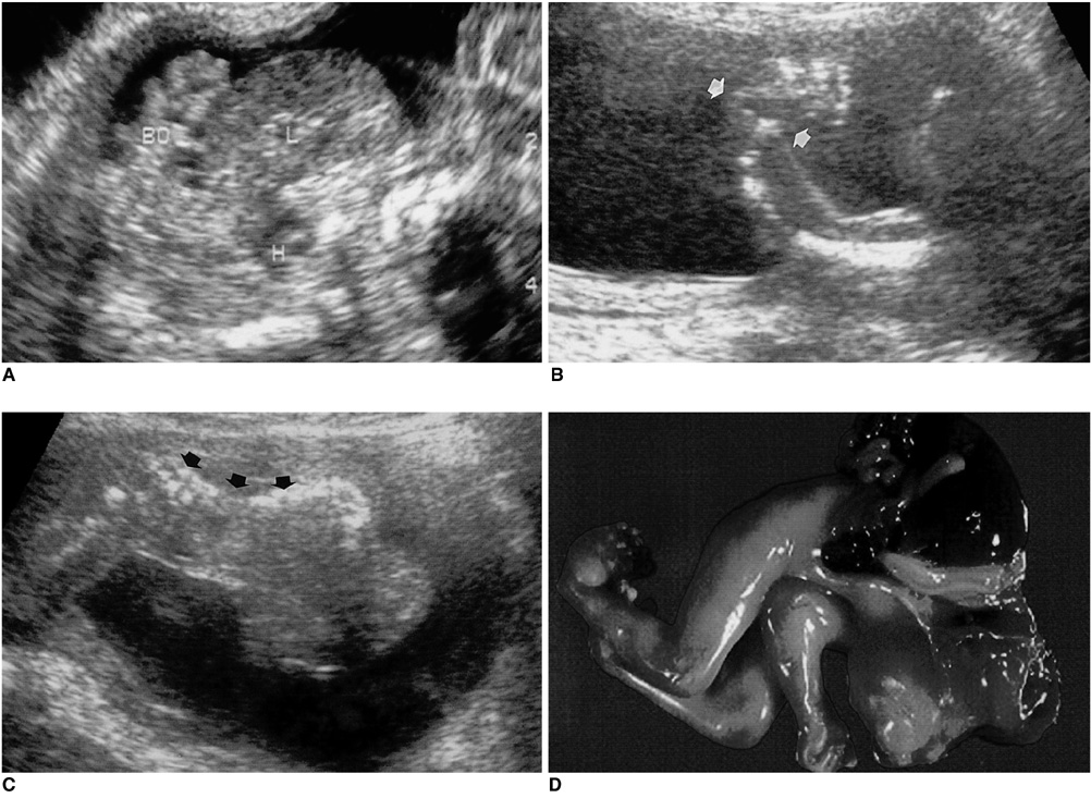
Fig. 10 Limb-body wall complex in a 14-week fetus. A. A complex mass consisting of the liver (L), bowel (BO), and heart (H) is identifiable lateral to the fetal abdomen and thorax at transvaginal ultrasonography. B. The ankle (arrows) is abnormally angulated. C. Kyphoscoliotic spinal curvature is apparent (arrows). D. Postmortem photograph demonstrates the presence of limb-body wall complex. Thoraco-abdominoschisis, limb anomalies, and exencephaly are present.

Fig. 11 Sirenomelia in a 16-week fetus. A. Prenatal ultrasonogram shows a single femur and tibia at the midline, and a deformed foot (arrowheads). The iliac bones (arrows) are abnormally located. B. Postmortem radiograph shows a single lower extremity and hypoplastic pelvic bone.
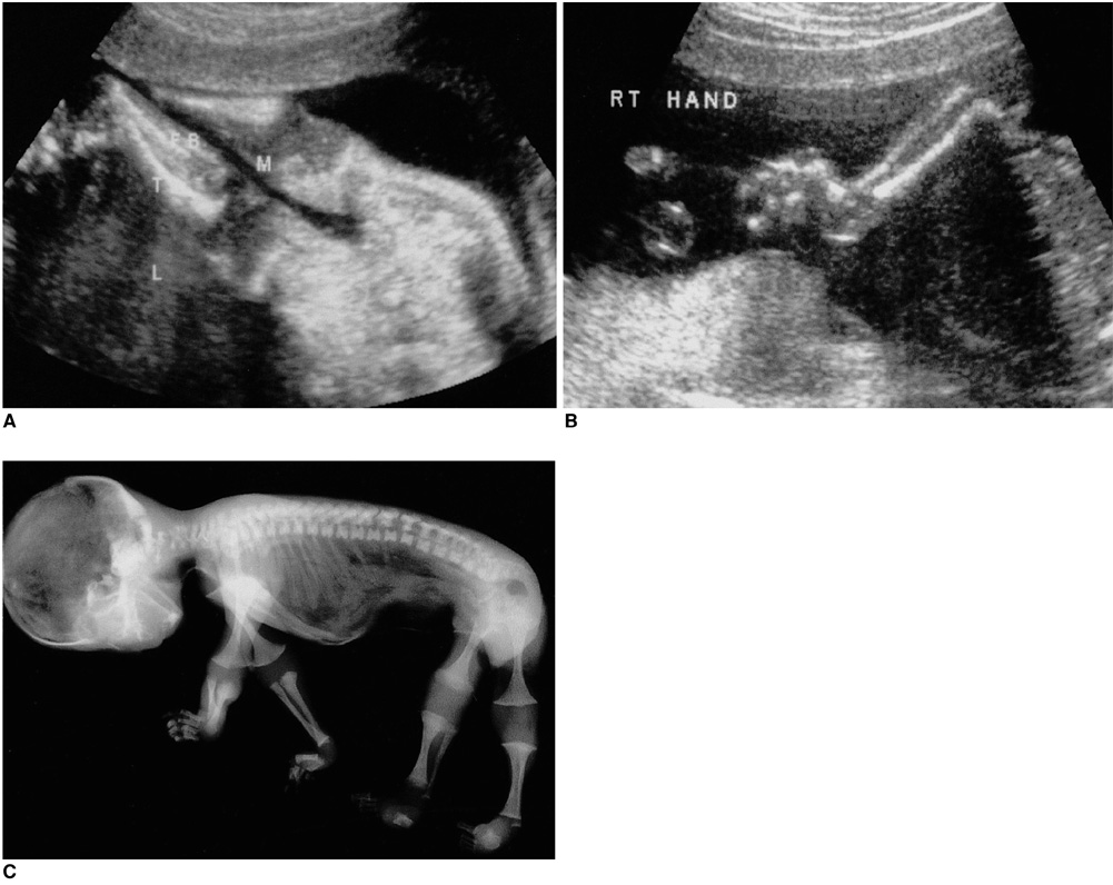
Fig. 12 Arthrogryposis multiplex congenita in a 19-week fetus. A. Ultrasonogram demonstrates unusual extension of both knees, without postural change. B. Both hands and fingers show contracture and abnormal angulation deformities. C. Postmortem radiograph shows extension deformity of the knee joints, clubfoot, and abnormal angulation deformity of both hands.
Reference
-
1. Stoll C, Dott B, Roth MP, Alembik Y. Birth prevalence rates of skeletal dysplasias. Clin Genet. 1989. 35:88–92.2. Maroteaux P, Lamy M, Rober JM. Le nanisme thanatophore. Presse Medicale. 1967. 75:2519–2524.3. Langer LO, Yang SS, Hall JG, et al. Thanatophoric dysplasia and cloverleaf skull. Am J Med Genet. 1987. 3:S. 167–179.4. Lemyre E, Azouz M, Teebi AS, Glanc P, Chen MF. Achondroplasia, hypophosphatasia and thanatophoric dysplasia: review and update. Can Assoc Radiol J. 1999. 50:185–197.5. Sillence DO, Barlow KK, Garber AP, Hall JG, Rimoin DL. Osteogenesis imperfecta type II: delineation of the phenotype with reference to genetic heterogeneity. Am J Med Genet. 1984. 17:407–423.6. Woo JSK, Ghosh A, Liang ST, et al. Ultrasonic evaluation of osteogenesis imperfecta congenita in utero. J Clin Ultrasound. 1983. 11:42–44.7. Whitley CB, Gorlin RJ. Achondrogenesis: new nosology with evidence of genetic heterogeneity. Radiology. 1983. 148:693–698.8. Mahony BS, Filly RA, Cooperberg PL. Antenatal sonographic diagnosis of achondrogenesis. J Ultrasound Med. 1984. 3:333–335.9. Tongsong T, Srisomboon J, Sudasna J. Prenatal diagnosis of Langer-Saldino achondrogenesis. J Clin Ultrasound. 1995. 23:56–58.10. Beighton P, Giedion A, Gorlin R, et al. International classification of osteochondrodysplasias. Eur J Pediatr. 1992. 151:407–415.11. Jones KL. Smith's Recognizable Patterns of Human Malformation. 1997. Philadelpia: Saunders.12. Naumoff P, Young LW, Mazer J, Amortegui AJ. Short rib-polydactyly syndrome type III. Radiology. 1977. 122:443–447.13. Beemer FA, Langer LO Jr, Klepde Pater JM, et al. A new short rib syndrome: report of two cases. Am J Med Genet. 1983. 14:115–128.14. Elejadle BR, de Elejadle MM, Pansch D. Prenatal diagnosis of Jeune syndrome. Am J Med Genet. 1985. 21:433–438.15. Ellis RWB, van Creveld S. A syndrome characterized by ectodermal dysplasia, polydactyly, chondrodysplasia and congenital morbus cordis. Arch Dis Child. 1940. 15:65.16. Cordone M, Lituania M, Zampatti C, et al. In utero ultrasonographic features of camptomelic dysplasia. Prenatal Diagn. 1989. 9:745–750.17. Lazjuk GI, Shved IA, Cherstvoy ED, Feshchenko SP. Camptomelic syndrome: Concepts of the bowing and shortening in the lower limbs. Teratology. 1987. 35:1–8.18. Spranger JW, Opitz JM, Bider U. Heterogeneity of chondrodysplasia punctata. Hum Genet. 1971. 11:190–212.19. Duff P, Harlass FE, Milligan DA. Prenatal diagnosis of chondrodysplasia punctata by sonography. Obstet Gynecol. 1990. 76:497–500.20. Torpin R. Amniochorionic mesoblastic fibrous strings and amniotic bands. Am J Obstet Gynecol. 1965. 91:65–75.21. Higginbottom MC, Jones KL, Hall BD, Smith DW. The amniotic disruption complex: timing of amniotic rupture and variable spectra of consequent defects. J Pediatr. 1979. 95:544–549.22. Burton DJ, Filly RA. Sonographic diagnosis of the amniotic band syndrome. AJR. 1991. 158:555–558.23. Van Allen MI, Curry C, Gallagher L. Limb-body wall complex: I. Pathogenesis. Am J Med Genet. 1987. 28:529–548.24. Balentyne G, Moesinger AC, James LS, Blank WA. Short umbilical cord and multiple anomalies in experimental oligohydramnios. Teratology. 1978. 17:S2. 43A.25. Patten RM, Van Allen M, Mack LA, et al. Limb-body wall complex: In utero sonographic diagnosis of a complicated fetal malformation. AJR. 1986. 146:1019–1024.26. Stevenson RE, Jones KL, Phelan MC, et al. Vascular steal: the pathogenetic mechanism producing sirenomelia and associated defects of the viscera and soft tissues. Pediatrics. 1986. 78:451–457.27. Sirtori M, Ghidini A, Romero R, Hobbins JC. Prenatal diagnosis of sirenomelia. J Ultrasound Med. 1989. 8:83–88.28. Sepulveda W, Romero R, Pryde PG, Wolfe HM, Addis JR, Cotton DB. Prenatal diagnosis of sirenomelus with color Doppler ultrasonography. Am J Obstet Gynecol. 1994. 170:1377–1379.29. Hall JG. Arthrogryposis multiplex congenita: etiology, genetics, classification, diagnostic approach, and general aspects. J Pediatr Orthop B. 1997. 6:159–166.30. Thompson G, Bilenker R. Comprehensive management of arthrogryposis multiplex congenita. Clin Orthop Relat Res. 1985. 194:6–14.31. Kirkinrn P, Herva R, Leisti J. Early prenatal diagnosis of a lethal syndrome of multiple congenital contractures. Prenat Diagn. 1987. 7:189–196.
- Full Text Links
-
- Actions
-
Cited
- CITED
-
- Close
- Share
-
- Similar articles
-
- The Usefulness of Ultrasonographic Evaluation in the Musculoskeletal Disease
- Prenatal Diagnosis and Clinical Outcome of Fetal Ovarian Cysts
- The predictive value of abnormal ultrasonographic finding for fetal trisomy in the second trimester
- Prenatal Ultrasound Findings of Fetal Neoplasms
- Ultrasonographic findings of early abortion: suggested predictors
