Focal Nodular Hyperplasia with Retraction of Liver Capsule: A Case Report
- Affiliations
-
- 1Department of Diagnostic Radiology, Kyung Hee University Hospital, Seoul, Korea. donghlee@hananet.net
- 2Department of Pathology, Kyung Hee University Hospital, Seoul, Korea.
- KMID: 754045
- DOI: http://doi.org/10.3348/kjr.2003.4.1.66
Abstract
- Focal nodular hyperplasia (FNH) is characterized by the presence a central scar with radiating fibrous septa. Our case had a capsular retraction, which was the result of an extension of the central scar to the surface. In addition, a hypointense scar on the T2-weighted image and a minimal enhancing central scar on the enhanced T1-weighted image, which was due to dense, sclerotic collagenous tissue, were observed. We report the first case of FNH with a capsular retraction.
Figure
-
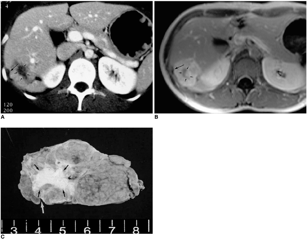
Fig. 1 A. 28-year-old woman with focal nodular hyperplasia with a retraction of the liver capsule. On hepatic helical CT scan during the portal phase, the mass shows an iso-attenuation with the liver parenchyma. The mass has a central fibrotic scar (small black arrows), and the liver capsule adjacent to the mass is retracted (large arrow). B. Contrast enhanced T1-weighted MR image reveals that the tumor is well enhanced with a minimally enhancing central scar (small black arrows). The lateral surface of the liver parenchyma adjacent to the mass is retracted (large arrow). C. The hepatic mass of segment 6 is measures approximately 3.5×5.5 cm. The cut surface of the mass reveals a nodular configuration with a central fibrous scar, extending to the liver surface (white arrow). The resected specimen shows a depressed thickened stellate scar (black arrows), slightly eccentrically positioned, with tapering fibrous septa that radiate through the mass, dividing it into multiple lobules.
Reference
-
1. Kehagias D, Moulopulos L, Antoniou A, et al. Focal nodular hyperplasia: imaging findings. Eur Radiol. 2001. 11:202–212.2. Mortele K J, Praet M, Van Vlierberghe H, Kunnen M. CT and MR imaging in focal nodular hyperplasia of the liver: Radiologic-Patholgic Correlation. AJR Am J Roentgenol. 2001. 175:687–692.3. Soyer P, Blumke DA, Vissuzaine C, Van Beers B, Barge J, Levesque M. CT of hepatic tumors : prevalence and specificity of retraction of the adjacent liver capsule. AJR Am J Roentgenol. 1994. 162:1119–1122.4. Seo BK, Rhee JY, Seol HY, Lee KY, Park CM, Chung KB. CT features of malignant hepatic tumors : The significance of capsular retraction. J Korean Radiol Soc. 1998. 38:267–271.5. Miller WJ, Dodd GD III, Federle MP, Baron RL. Epitheloid hamangioendothelioma of the liver: Imaging findings with pathologic correlation. AJR Am J Roentgenol. 1992. 159:53–57.6. Shamsi K, De Schepper A, Degryse H, Deckers F. Focal nodular hyperplasia of the liver: radiologic findings. Abdom Imaging. 1993. 18:32–38.7. Vilgrain V, Flejou J, Arrive L, et al. Focal nodular hyperplasia of the liver: MR imaging and pathologic correlation in 37 patients. Radiology. 1992. 184:699–703.8. Rummeny E, Weissleder R, Sironi S, et al. Central scars in primarily liver tumors: MR features, specificity, and pathologic correlation. Radiology. 1989. 171:323–326.9. Yang DM, Yoon MH, Kim HS, Chung JW. Capsular5 retraction in hepatic hemangioma : CT and MR features. Abdom Imaging. 2001. 26:36–38.10. Blachar A, Federle MP, Brancatelli G. Hepatic capsular retraction: spectrum of benign and malignant etiologies. Abdom Imaging. 2002. 27:690–699.
