Differential Diagnosis of Benign and Malignant Intraductal Papillary Mucinous Tumors of the Pancreas: MR Cholangiopancreatography and MR Angiography
- Affiliations
-
- 1Department of Radiology, Asan Medical Center, University of Ulsan College of Medicine. tkkim@amc.seoul.kr
- 2Department of Surgery, Asan Medical Center, University of Ulsan College of Medicine.
- KMID: 754019
- DOI: http://doi.org/10.3348/kjr.2003.4.3.157
Abstract
OBJECTIVE
To compare the usefulness of magnetic resonance cholangiopancreatography (MRCP) and MR angiography (MRA) in differentiating malignant from benign intraductal papillary mucinous tumors of the pancreas (IPMTs), and to determine the findings which suggest malignancy. MATERIALS AND METHODS: During a 6-year period, 46 patients with IPMT underwent MRCP. Morphologically, tumor type was classified as main duct, branch duct, or combined. The diameter of the main pancreatic duct (MPD), the extent of the dilated MPD, and the location and size of the cystic lesion, septum, and communicating channel were assessed. For all types of IPMTs, enhanced mural nodules and portal vein narrowing were evaluated at MRA. RESULTS: Combined-type IPMTs were more frequently malignant (78%) than benign (42%) (p < 0.05). Compared with benign lesions, malignant lesions were larger, and the caliber of the communicating channel was also larger (p < 0.05). Their dilated MPD was more extensive and of greater diameter (p < 0.05), and the presence of mural nodules was more frequent (p < 0.001). CONCLUSION: Combined MRCP and MRA might be useful for the differential diagnosis of malignant and benign IPMTs of the pancreas.
Figure
-

Fig. 1 A 60-year-old man with benign main duct-type IPMT of the pancreas. Single-slab MRCP image (TR/TE, infinite/1200) shows diffuse dilatation of the main pancreatic duct (arrows).
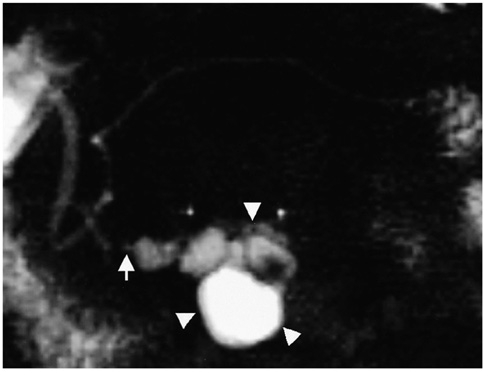
Fig. 2 A 44-year-old man with benign branch duct-type IPMT of the pancreas. Single-slab MRCP image (TR/TE, infinite/1200) depicts a multilocular cystic lesion (arrowheads) in the pancreatic uncinate process and a narrow channel (arrow) which communicates with the pancreatic duct.
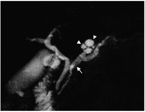
Fig. 3 A 67-year-old man with benign combined duct-type IPMT of the pancreas. Single-slab MRCP image (TR/TE, infinite/1200) reveals a multilocular cystic lesion (arrowheads) in the pancreatic body and diffuse mild dilation of the main pancreatic duct (arrow).
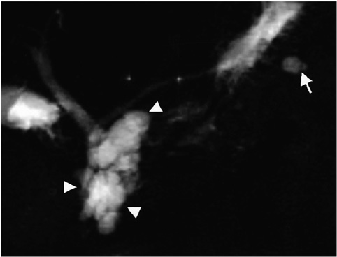
Fig. 4 A 64-year-old woman with malignant branch duct-type IPMT of the pancreas. Single-slab MRCP image (TR/TE, infinite/1200) demonstrates a grapelike cluster of cysts (arrowheads). The main pancreatic duct is not dilated. Another small cystic lesion (arrow), not resected at surgery, is seen in the pancreatic tail.
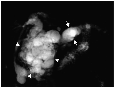
Fig. 5 A 71-year-old man with malignant combined duct-type IPMT of the pancreas. Single-slab MRCP image (TR/TE, infinite/1200) depicts a grapelike cluster of cysts (arrowheads) in the pancreatic head. Marked diffuse dilation of the main pancreatic duct is observed (arrows).

Fig. 6 A 44-year-old man with malignant combined duct-type IPMT of the pancreas. The MRA source image (TR/TE, 4.6/1.8) shows an enhancing mural nodule (arrow) and a thick irregular septum in the multilocular cystic lesion (arrowheads) of the pancreatic head.
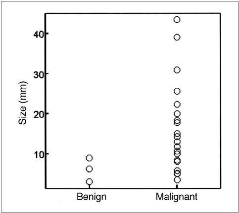
Fig. 7 Scatterplot showing the size of mural nodules in benign and malignant IPMTs of the pancreas. Some overlap is apparent, but in benign IPMTs the mural nodule is less than 10 mm in diameter.
Reference
-
1. Yamada M, Kozuka S, Yamao K, Nakazawa S, Naitoh Y, Tsukamoto Y. Mucin-producing tumor of the pancreas. Cancer. 1991. 68:159–168.2. Doi R, Fujimoto K, Wada M, Imamura M. Surgical management of intraductal papillary mucinous tumor of the pancreas. Surgery. 2002. 132:80–85.3. Kimura W, Sasahira N, Yoshikawa T, Muto T, Makuuchi M. Duct-ectatic type of mucin producing tumor of the pancreas-new concept of pancreatic neoplasia. Hepatogastroenterology. 1996. 43:692–709.4. Sugiyama M, Atomi Y. Intraductal papillary mucinous tumors of the pancreas: imaging studies and treatment strategies. Ann Surg. 1998. 228:685–691.5. Yamaguchi K, Ogawa Y, Chijiiwa K, Tanaka M. Mucin-hypersecreting tumors of the pancreas: assessing the grade of malignancy preoperatively. Am J Surg. 1996. 171:427–431.6. Uehara H, Nakaizumi A, Iishi H, et al. Cytologic examination of pancreatic juice for differential diagnosis of benign and malignant mucin-producing tumors of the pancreas. Cancer. 1994. 74:826–833.7. Sugiyama M, Atomi Y, Kuroda A. Two types of mucin-producing cystic tumors of the pancreas: diagnosis and treatment. Surgery. 1997. 122:617–625.8. Sugiyama M, Atomi Y, Hachiya J. Intraductal papillary tumors of the pancreas: evaluation with magnetic resonance cholangiopancreatography. Am J Gastroenterol. 1998. 93:156–159.9. Koito K, Namieno T, Ichimura T, et al. Mucin-producing pancreatic tumors: comparison of MR cholangiopancreatography with endoscopic retrograde cholangiopancreatography. Radiology. 1998. 208:231–237.10. Onaya H, Itai Y, Niitsu M, Chiba T, Michishita N, Saida Y. Ductectatic mucinous cystic neoplasm of the pancreas: evaluation with MR cholangiopancreatography. AJR Am J Roentgenol. 1998. 171:171–177.11. Fukukura Y, Fujiyoshi F, Sasaki M, et al. HASTE MR cholangiopancreatography in the evaluation of intraductal papillary-mucinous tumors of the pancreas. J Comput Assist Tomogr. 1999. 23:301–305.12. Irie H, Honda H, Aibe H, et al. MR cholangiopancreatographic differentiation of benign and malignant intraductal mucin-producing tumors of the pancreas. AJR Am J Roentgenol. 2000. 174:1403–1408.13. Kim TK, Kim BS, Kim JH, et al. Diagnosis of intrahepatic stones: superiority of MR cholangiopancreatography over endoscopic retrograde cholangiopancreatography. AJR Am J Roentgenol. 2002. 179:429–434.14. Kim JH, Kim TK, Eun HW, et al. Preoperative evaluation of gallbladder carcinoma: efficacy of combined use of MR imaging, MR cholangiography, and contrast-enhanced dual-phase three-dimensional MR angiography. J Magn Reson Imaging. 2002. 16:676–684.15. Kobayashi G, Fujita N, Lee S, Kimura K, Watanabe H, Mochizuki F. Correlation between ultrasonographic findings and pathological diagnosis of mucin-producing tumor of the pancreas. Nippon Shokakibyo Gakkai Zasshi. 1990. 87:235–242.16. Yanagisawa A, Ohashi K, Hori M, et al. Ductectatic-type mucinous cystadenoma and cystadenocarcinoma of the human pancreas: a novel clinicopathological entity. Jpn J Cancer Res. 1993. 84:474–479.
- Full Text Links
-
- Actions
-
Cited
- CITED
-
- Close
- Share
-
- Similar articles
-
- Intraductal Papillary Mucinous Tumor of the Pancreas: Usefulness of Endoscopic Ultrasonography in Differentiation of Benign and Malignant Neoplasm
- Cystic Neoplasms and Intraductal Papillary Mucinous Neoplasms of the Pancreas
- Evaluation of malignant intraductal papillary mucinous neoplasms of the pancreas on computed tomography and magnetic resonance imaging
- Comparison of Mucinous Cystic Tumor and Intraductal Papillary Mucinous Tumor
- A Case of Intraductal Papillary Mucinous Neoplasm Arising from Santorini's Duct in a Patient with Complete Type of Pancreas Divisum
