Korean J Radiol.
2004 Jun;5(2):139-142. 10.3348/kjr.2004.5.2.139.
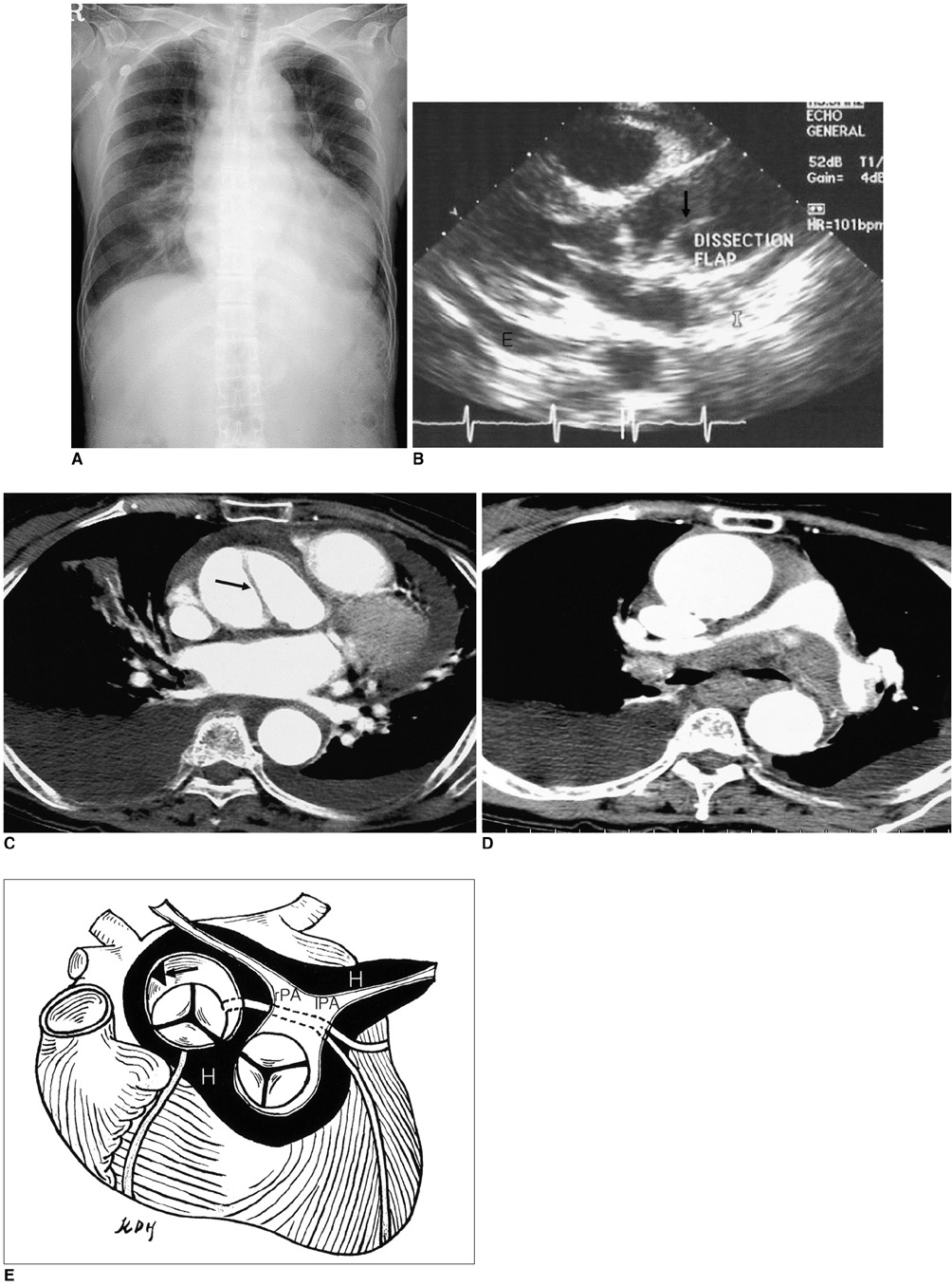
Aortic Dissection Presenting with Secondary Pulmonary Hypertension Caused by Compression of the Pulmonary Artery by Dissecting Hematoma: A Case Report
- Affiliations
-
- 1Department of Radiology, Soonchunhyang University Hospital. dhk1107@hanmail.net
- 2Department of Thoracic and Cardiovascular Surgery, Chonnam National University Hospital.
- KMID: 753995
- DOI: http://doi.org/10.3348/kjr.2004.5.2.139
Abstract
- The rupture of an acute dissection of the ascending aorta into the space surrounding the pulmonary artery is an uncommon occurrence. No previous cases of transient pulmonary hypertension caused by a hematoma surrounding the pulmonary artery have been documented in the literature. Herein, we report a case of acute aortic dissection presenting as secondary pulmonary hypertension.
Keyword
MeSH Terms
Figure
Reference
-
1. Kahn SE, Kotler MN, Goldman AP, Ablaza S. Superior vena caval obstruction secondary to acute dissecting aneurysm of the aorta. Am Heart J. 1986. 111:606–608.2. Rau AN, Glass MN, Waller BF, Fraiz J, Shaar CJ. Right pulmonary artery occlusion secondary to a dissecting aortic aneurysm. Clin Cardiol. 1995. 18:178–180.3. Buja LM, Ali N, Fletcher RD, Roberts WC. Stenosis of the right pulmonary artery: a complication of acute dissecting aneurysm of the ascending aorta. Am Heart J. 1972. 83:89–92.4. Spier LN, Hall MH, Nelson RL, Parnell VA, Pogo GJ, Tortolani AJ. Aortic dissection: rupture into right ventricle and right pulmonary artery. Ann Thorac Surg. 1995. 59:1017–1019.5. Charnsangavej C. Occlusion of the right pulmonary artery by acute dissecting aortic aneurysm. AJR Am J Roentgenol. 1979. 132:274–276.6. Castaner E, Andreu M, Gallardo X, Mata FM, Cabezuelo MA, Pallardo Y. CT in nontraumatic acute thoracic aortic disease: typical and atypical features and complications. RadioGraphics. 2003. 23:S93–S110.7. Nasrallah A, Goussous Y, El-Said G, Garcia E, Hall RJ. Pulmonary artery compression due to acute dissecting aortic aneurysm: clinical and angiographic diagnosis. Chest. 1975. 67:228–230.8. Fattori R. Higgins CB, de Roos A, editors. MRI and MRA of thoracic aorta. Cardiovascular MRI & MRA. 2003. Philadelphia: Lippincott Williams & Wilkins;375–386.9. Dzau VJ, Creager MA. Isselbacher KJ, Braunwald E, Wilson JD, Martin JB, Fauci AS, Kasper DL, editors. Diseases of the aorta. Harrison's principles of internal medicine. 1992. Vol 1:13th ed. New York: McGraw-Hill, Inc;1131–1135.
- Full Text Links
- Actions
-
Cited
- CITED
-
- Close
- Share
- Similar articles
-
- Pulmonary Artery Periadventitial Hematoma in a Patient with Aortic Intramural Hematoma: A Case Report
- A Case of Extrinsic Compression of the Left Main Coronary Artery Secondary to Pulmonary Artery Dilatation
- A Dissecting Aneurysm of the Common and Proper Hepatic Artery with Dissection of the Celiac Axis and the SMA
- Differential Imaging Features of Pulmonary Artery Dissection from Other Intraluminal Diseases of Pulmonary Artery: Two Cases Report
- A Case of Internal Carotid Artery Dissection Presenting with Isolated Hypoglossal Nerve Palsy


