Yonsei Med J.
2008 Jun;49(3):509-513. 10.3349/ymj.2008.49.3.509.
Atypical Angioma Serpiginosum
- Affiliations
-
- 1Department of Dermatology, Taipei Municipal Wan-Fang Hospital, Taipei Medical University, Taipei, Taiwan. shiunkle @mail2000.com.tw
- 2Department of Dermatology, Taipei Medical University Hospital, Taipei, Taiwan.
- 3Medical Imaging Lab and Medical Intelligence Network, Department of Nuclear Medicine, Buddhist Dalin Tzu Chi General Hospital, Chiayi, and Department of Medicine, College of Medicine, Tzu Chi University, Hualien, Taiwan.
- KMID: 724271
- DOI: http://doi.org/10.3349/ymj.2008.49.3.509
Abstract
- Angioma serpiginosum is an uncommon, acquired vascular nevoid disorder with capillary dilation and proliferation in the papillary dermis. The eruptions are asymptomatic and characterized by grouped, erythematous to violaceous, serpiginous and punctate macules. The condition usually appears in females during adolescence on unilateral lower extremities and the buttocks. We report a rare case with a late onset and atypical distribution of lesions in a 48-year-old female patient who had groups of punctate lesions on her left foot for four to five years. Histopathological examination showed hyperkeratosis and multiple dilated and proliferated capillaries in the papillary dermis. Inflammation and extravasation of red blood cells were not found. According to the clinical and pathological findings, we established a diagnosis of angioma serpiginosum. She was treated with a pulsed dye laser, and the angiomatous lesions subsequently improved.
Keyword
MeSH Terms
Figure
-
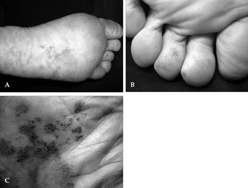
Fig. 1 (A) Grouped, punctate, erythematous macules on the left sole. (B) Similar lesions on toes. (C) Close-up view of the lesions on left sole.

Fig. 2 (A) Hyperkeratosis and many dilated and proliferated capillaries in the papillary dermis (hematoxylin and eosin, original magnification × 100). (B) Dilated and proliferated congested capillaries at higher power (hematoxylin and eosin, original magnification × 200).
Reference
-
1. Cox NH, Paterson WD. Angioma serpiginosum: a simulator of purpura. Postgrad Med J. 1991. 67:1065–1066.
Article2. Al Hawsawi K, Al Aboud K, Al Aboud D, Al Githami A. Linear angioma serpiginosum. Pediatr Dermatol. 2003. 20:167–168.
Article3. Namazi MR, Handjani F. Angioma serpiginosum. Dermatol Online J. 2003. 9:19.
Article4. Hunt SJ, Santa Cruz DJ. Acquired benign and "borderline" vascular lesions. Dermatol Clin. 1992. 10:97–115.5. Ohnishi T, Nagayama T, Morita T, Miyazaki T, Okada H, Ohara K, et al. Angioma serpiginosum: a report of 2 cases identified using epiluminescence microscopy. Arch Dermatol. 1999. 135:1366–1368.6. Barker LP, Sachs PM. Angioma serpiginosum, a comparative study. Arch Dermatol. 1965. 92:613–620.
Article7. Marriott PJ, Munro DD, Ryan T. Angioma serpiginosum-familial incidence. Br J Dermatol. 1975. 93:701–706.
Article8. Katta R, Wagner A. Angioma serpiginosum with extensive cutaneous involvement. J Am Acad Dermatol. 2000. 42:384–385.
Article9. Gautier-Smith PC, Sanders MD, Sanderson KV. Ocular and nervous system involvement in angioma serpiginosum. Br J Ophthalmol. 1971. 55:433–443.
Article10. Erbagci Z, Erbagci I, Erkiliç S, Bekir N. Angioma serpiginosum with retinal involvement in a male: a possible aetiological role of continuous cold exposure. J Eur Acad Dermatol Venereol. 2004. 18:238–239.
Article11. Requena L, Sangueza OP. Cutaneous vascular proliferation. Part II. Hyperplasias and benign neoplasms. J Am Acad Dermatol. 1997. 37:887–919. quiz 920-2.12. Kumakiri M, Katoh N, Miura Y. Angioma serpiginosum. J Cutan Pathol. 1980. 7:410–421.
Article13. Xiao X, Hong L, Sheng M. Promoting effect of estrogen on the proliferation of hemangioma vascular endothelial cells in vitro. J Pediatr Surg. 1999. 34:1603–1605.
Article14. Neumann E. Some new observations on the genesis of angioma serpiginosum. Acta Derm Venereol. 1971. 51:194–198.15. Tok J, Berberian BJ, Sulica VI. Unilateral nevoid telangiectasia syndrome. Cutis. 1994. 53:53–54.16. Landthaler M, Haina D, Waidelich W, Braun-Falco O. A three-year experience with the argon laser in dermatotherapy. J Dermatol Surg Oncol. 1984. 10:456–461.17. Polla LL, Tan OT, Garden JM, Parrish JA. Tunable pulsed dye laser for the treatment of benign cutaneous vascular ectasia. Dermatologica. 1987. 174:11–17.18. Long CC, Lanigan SW. Treatment of angioma serpiginosum using a pulsed tunable dye laser. Br J Dermatol. 1997. 136:631–632.
Article19. Wheeland RG. Treatment of port-wine stains for the 1990s. J Dermatol Surg Oncol. 1993. 19:348–356.20. Spicer MS, Goldberg DJ. Lasers in dermatology. J Am Acad Dermatol. 1996. 34:1–25. quiz 26-8.
